Breast cancer: what false results reveal
All content is checked by medical journalists.Women over 50 are regularly invited to mammography in Germany. The screening is intended to detect breast cancer in the very early stages of the disease. But breast screening is coming under increasing criticism because false-positive findings - i.e. suspected cancer that later turns out to be irrelevant - often cause unnecessary fears. According to a new study, however, these incorrect findings could be associated with a higher risk of developing cancer later.
Women with false positive results were almost 40 percent more likely to develop breast cancer in the following ten years than women with normal mammography results. Women with a false-positive result who were then subjected to a biopsy even had a 76 percent higher risk.
“It is possible that suspicious findings, even if no cancer can be detected initially, are associated with later tumor formation,” explains Prof. Louise M. Henderson of the University of North Carolina.
The calculations were based on data from over two million mammograms from around 1.3 million women in the United States. They are based on the Breast Cancer Surveillance Consortium, which observed women for more than ten years between 1994 and 2009.
Positive finding in the mammography
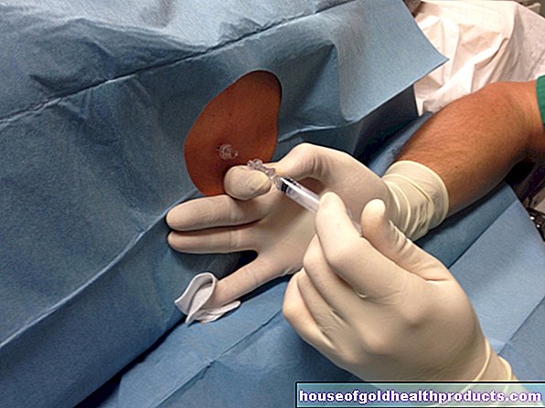
A conspicuous finding in a preventive examination is usually checked using other imaging methods, such as magnetic resonance imaging (MRI). If the suspicion of cancer cannot be refuted, a biopsy is performed and, in some cases, immediate surgery is recommended.
A total of almost 49,000 women were diagnosed with breast cancer during the observation period. However, on the basis of the data, the researchers were unable to determine whether the tumor had grown in the previously conspicuous one, in one place in the breast or even in the other breast.
Dense breast tissue as a risk factor
The female breast consists mainly of adipose and connective tissue. Embedded in it is the milk-producing glandular tissue, which consists of grape-shaped glandular lobules that are connected to the nipple via milk ducts. The breast tissue also contains nerves, veins and lymph vessels. If the tissue is particularly dense, the risk of breast cancer is higher than with less dense tissue.
The researchers therefore investigated whether the density of the breast tissue influenced the relationship between false-positive findings and future breast cancer diagnoses. More false positives were diagnosed in women with very dense breast tissue than in women with less dense breasts, which consisted primarily of adipose tissue. "We weren't surprised because the images of dense breast tissue are much more difficult to interpret," says Henderson. However, the density of the tissue had no influence on the likelihood of actually developing breast cancer after a false positive result.
False-positive results as a risk indicator
Henderson suggests using false-positive results in the future as an indicator of an increased risk of breast cancer and that this can be included in the calculation of individual breast cancer probabilities - similar to age, breast tissue density and cancer in the family history.
Why false-negative findings are more often associated with later cancer has not yet been clearly clarified. It is possible that the harmless changes in the pictures will later develop into a tumor more often.
However, the link may also be based on the fact that women who know they have an increased risk - for example, from having cancer in their family history - go to check-ups more often. This would automatically increase the likelihood that you will receive an incorrect result.
Positive result without breast cancer
The more often mammograms or other breast examinations are performed, the more often incorrect findings are made. Out of ten women women who have their breasts examined every two years, four receive a false-positive result within ten years. There are as many as six of the women who undergo annual examinations.
Microcalcifications or precursors to cancer can be falsely suspected of being malignant. Only the histological examination of the tissue can prove what the radiologists have seen in the images and found to be conspicuous.
The more often preventive examinations are carried out, the more often malignant tumors are found, which can then be treated. Whether and how often you want to have mammograms carried out therefore requires careful weighing of the benefits and risks. (vv)
Tags: elderly care healthy workplace smoking


.jpg)