Neurological examination
Valeria Dahm is a freelance writer in the medical department. She studied medicine at the Technical University of Munich. It is particularly important to her to give the curious reader an insight into the exciting subject area of medicine and at the same time to maintain the content.
More about the experts All content is checked by medical journalists.With the help of a neurological examination, the doctor checks the function and performance of the brain and nervous system. In addition to a thorough medical history and a detailed physical examination, special neurological tests help him. Read all about the neurological exam, how it works, and what the risks are.

What is a neurological exam?
Neurology deals with diseases of the nervous system. These include the brain and spinal cord (central nervous system, CNS), the cranial nerves, and the nerves that run through the entire body (peripheral nervous system, PNS). If the doctor suspects a disorder of the nervous system, he can often find the cause and the location of the complaint through a careful neurological examination. He checks the various functions of the nerves. The neurological exam includes:
- a medical discussion about the medical history and current complaints (anamnesis)
- a psychological finding about the patient's level of consciousness
- the palpation of the pulses and a blood pressure measurement
- the examination of the twelve cranial nerves
- the study of strength, sensitivity, reflexes and coordination of the body
- checking the stance, gait and balance
A balance test is important because dizziness and balance disorders are among the most common neurological symptoms. Special neurological tests such as the Unterberger stepping test, the Romberg standing test (Romberg test), the finger-nose test, the knee-heel test or the caloric test make diagnosis easier for the doctor. The right side is always compared with the left side in order to discover any deviations.
When to do a neurological exam
The neurological examination is the first step in diagnosing diseases of the nervous system. They often allow a good assessment of the cause and localization without having to initiate complex technical examinations or laboratory determinations. Common reasons for a neurological exam are:
- acute circulatory disorders in the CNS, e.g. in the case of a stroke
- Brain tumors or abscesses that displace healthy tissue in the cranial cavity and thereby cause discomfort
- Herniated discs
- Epilepsies
- chronic inflammatory diseases of the CNS, e.g. multiple sclerosis
- acute inflammation of the brain or meninges
- Metabolic disorders of the peripheral nerves, e.g. due to diabetes (diabetic polyneuropathy)
- pressure-related dysfunction of the peripheral nerves
- dizziness
What do you do during a neurological exam?
At the beginning, the doctor assesses the patient's wakefulness (vigilance), for example by asking for the date of birth, first name or whereabouts. If the patient can answer everything correctly, his condition is classified as "awake and oriented". In addition, the previous medical history and the current complaints are recorded. The blood pressure is measured and the pulse is palpated.
In addition, the doctor checks the sensitivity of the entire body. Sensations of touch, pain, temperature, vibration and changes in position are tested. He also examines motor skills and divides muscle strength into different strength levels. In this way, paralysis or cramps (spasticity) can be recognized.
The neurological examination of coordination can be carried out using the finger-nose experiment. The eyes are closed and the index finger of the outstretched arm is brought to the tip of the nose. The knee-heel attempt is an alternative. Stance, gait and balance can be checked using the Romberg standing test and the Unterberger step test. You should take 50 steps with your eyes closed on the spot, without turning too much.
Review of the cranial nerves
The cranial nerves, which arise directly from the brain, are examined separately from each other in the neurological examination:
- I. Olfactory nerve - Smell: Verification by smell tests
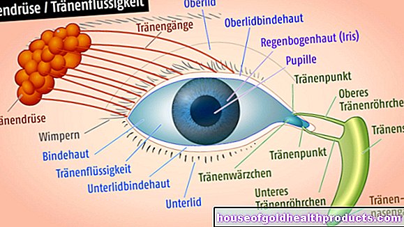
- II. Optic nerve - vision: Objects or letters must be recognized from a certain distance. The pupil response is checked by the doctor shining a lamp in the eyes and assessing the pupillary response.
- III. Oculomotor nerve - eye movement: Here the patient should be able to follow the doctor's finger with his eyes
- IV. Trochlear Nerve - Eye Movement: The patient looks inward and downward for verification. The doctor tests both eyes separately.
- V. trigeminal nerve - chewing and sensitivity: the doctor strokes the patient's face and asks if he can feel the touch. It also presses on the exit points of the nerves above the eyebrows, below the eyes and on the chin. This shouldn't cause pain.
- VI. Abducens nerve - eye movement: the patient looks outward to check. Here, too, tests are carried out in a side-by-side comparison.
- VII. Facial nerve - facial expressions and taste: Here the patient inflates his cheeks, frowns and makes a kiss. In addition, the patient's taste perception is asked about.
- VIII. Vestibulocochlear Nerve - Hearing and Balance: The doctor rubs fingers near the ears to check hearing. The nerve function is checked with a balance test.
- IX. Glossopharyngeal Nerve - Swallowing: The doctor inspects the throat and the ability to swallow
- X. Vagus nerve - control of internal organs: the doctor asks about abnormalities in the heartbeat, breathing or digestion
- XI. Accessor nerve - part of the head muscles: the doctor pushes the shoulders down while the patient pulls them up. In addition, the head should be able to be turned against resistance.
- XII.Hypoglossal nerve - tongue: the patient sticks out the tongue and moves it to all sides
To rule out meningitis and other diseases, the patient places his chin on his chest. If pain occurs here, one speaks of meningism (neck stiffness), which must be examined more closely.
Examination of the reflexes
The neurological examination also includes the examination of the reflexes. With the help of a reflex hammer, the doctor tests the so-called muscle reflexes such as the biceps tendon reflex. The doctor puts a thumb on the biceps tendon and hits it with the hammer. If the forearm bends, injuries to the nerves involved are almost impossible.
With the so-called external reflexes, the reflex response does not take place in the stimulus-perceiving organ. If the doctor wipes the thigh, for example, the man should lift the testicle.
In addition, the primitive reflexes are tested, which should no longer be triggered in healthy people and are only present in newborns and small children. With the Babinski reflex, for example, the outer edge of the foot is painted vigorously. If there is nerve damage, the toes spread apart and the big toe lifts up.
What are the risks of a neurological examination?
A neurological examination is a complicated but uncomplicated examination. Injuries such as bruises (bruises), wounds or nerve, muscle and soft tissue damage can occur in rare cases if the doctor uses too much force during the examination - for example, by striking too hard with the reflex hammer. During the balance test, the patient should be protected in the event that he becomes unbalanced.
What do I have to consider after a neurological examination?
Once the neurological exam is complete, your doctor will discuss the results with you. Depending on the diagnosis, further technical neurological examinations such as magnetic resonance tomography (MRT), computed tomography (CT) or electroneurography (ENG) are carried out.
Tags: fitness parasites prevention