Aortic stenosis
Dr. med. Julia Schwarz is a freelance writer in the medical department.
More about the experts All content is checked by medical journalists.Aortic valve stenosis is the most common heart valve defect that requires treatment. The cause is usually acquired calcification. The oxygen-rich blood can no longer be adequately pumped into the large circulation. Symptoms of aortic valve stenosis are an insufficient supply of the brain, dizziness and reduced exercise capacity. Read everything you need to know about aortic stenosis here.
ICD codes for this disease: ICD codes are internationally recognized codes for medical diagnoses. They can be found, for example, in doctor's letters or on certificates of incapacity for work. I08Q23I35I06
Aortic stenosis: description
Aortic valve stenosis (aortic stenosis) is the heart valve defect that requires treatment most often. Looking at the number of cases alone, mitral valve regurgitation is the most common heart valve defect in Europe and North America. However, it does not have to be treated as often as the aortic valve stenosis.
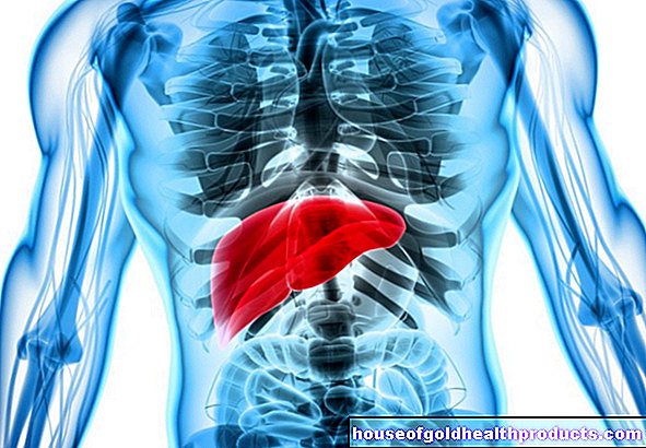
The aortic valve consists of three crescent-shaped pockets. It lies between the left ventricle and the main artery (aorta). It serves as a valve there so that the blood can only flow in one direction - namely into the large bloodstream - and does not flow back into the heart.
This “exit” from the heart is narrowed in aortic valve stenosis. Because of this resistance, the heart has to exert more force to open the valve and keep pumping the blood. As a result, the heart muscle thickens noticeably (hypertrophy). In the course of time it becomes less and less flexible and weaker, the pumping performance decreases. In the case of advanced aortic valve stenosis in particular, the muscle ultimately no longer manages to transport sufficient oxygen-rich blood into the body's circulation.
Aortic stenosis: symptoms
Mild or moderate aortic valve stenosis does not cause any symptoms for a long time. This is especially the case when those affected hardly exert themselves physically. Because the first symptoms appear under stress.
At the beginning, those affected usually complain of dizziness and an occasional circulatory collapse up to loss of consciousness (syncope). This is due to the lack of blood flow to the brain as a result of the aortic stenosis. The heart can hardly keep up, especially in situations of physical stress (climbing stairs or sports): because of the aortic valve stenosis, the heart can no longer pump enough blood out of the heart to meet the body's increased oxygen requirements during physical activity.
In order to pump against the aortic valve stenosis, the left ventricle needs more muscle strength. Over time, it adapts to this by enlarging (concentric left heart hypertrophy). The increase in myocardial tissue also increases its need for oxygen. In addition, the thickened muscle constricts the coronary arteries, which supply the heart with blood and oxygen, especially when it is tense. Patients complain of tightness or pain in the chest (angina pectoris), even when the coronary arteries themselves are healthy.
The insufficient supply of the heart muscle and its enlargement can ultimately lead to symptoms of cardiac insufficiency (heart failure) and / or to cardiac arrhythmias (for example atrial fibrillation). If the blood can no longer be pumped out of the heart properly, it backs up from the left ventricle back towards the lungs. If this stagnation pressure in the vessels continues to rise, liquid escapes and is deposited in the tissue. With increasing weakness of the heart, fluid eventually collects in the lung tissue (pulmonary edema), and patients complain of severe shortness of breath.
Therefore, pay attention to the first signs of heart failure: Your performance decreases, you are quickly weakened and you feel short of breath when you exert yourself. In addition, some symptoms start at night, such as a cough.
Aortic stenosis: causes and risk factors
Aortic stenosis can be acquired or congenital.
Acquired aortic stenosis
The aortic valve stenosis is acquired in most cases, most often as a result of wear processes (calcification) in old age. This process is similar to that of arteriosclerosis.Therefore, risk factors such as increased blood lipids promote the development of aortic valve stenosis. Lime and collagen are deposited in the valves. This thickens and hardens visibly. Initially referred to as aortic valve sclerosis, these processes eventually lead to a narrowing of the valve, which is why doctors then speak of aortic valve stenosis.
Rheumatic fever (which has become rare these days due to early, consistent antibiotic treatment of a streptococcal infection) can also cause scarring through autoimmune reactions and thus aortic valve stenosis: Scar tissue is less flexible than healthy tissue, which hinders the flow of blood from the heart into the aorta.
Congenital aortic stenosis
Congenital aortic valve stenosis is much rarer and causes symptoms even at a young age. It is a form of congenital aortic stenosis:
The heart valve itself is most frequently affected by the narrowing (valvular aortic valve stenosis). Usually it consists of only two pockets (bicuspid aortic valve). If it is not already constricted, two-leaved aortic valves stenose on average twenty years earlier than properly designed. If the area above the aortic valve (i.e. the beginning of the aorta) has narrowed, one speaks of a supravalvular aortic stenosis. With subvalvular aortic valve stenosis, the tissue below the heart valve is narrowed.
Aortic stenosis: examinations and diagnosis
If aortic valve stenosis is suspected, the doctor first asks about the medical history and possible complaints (anamnesis), for example:
- How active are you? (Sometimes symptoms of the aortic valve stenosis do not show up because the affected person hardly moves!)
- Do you feel more and more exhausted in the last few months?
- Do you tire quickly with physical exertion?
- Do you feel short of breath while doing this?
- Have you passed out lately?
- Do you have chest pain or pressure?
The doctor will then do a physical exam. He measures the patient's blood pressure (rather low), feels the pulse and listens to his heart with the stethoscope. In the case of aortic valve stenosis, a typical heart murmur can be heard during the ejection phase of the heartbeat (systolic). This heart murmur can usually still be heard at the level of the carotid arteries.
The doctor can hear the aortic valve stenosis best with a stethoscope between the second and third ribs directly to the right of the sternum.
In order to confirm the diagnosis of "aortic valve stenosis", an apparatus-based diagnosis is usually carried out afterwards:
roentgen
In the chest x-ray, the doctor can see any wall thickening of the left ventricle or enlargement of the aorta. A lateral x-ray can even show the calcification of the aortic valve.
Electrocardiography (EKG)
Usually, if aortic valve stenosis is suspected, an EKG is also performed. The thickening of the wall of the left ventricle is particularly evident in the typical jagged pattern.
Echocardiography
An echocardiography is an ultrasound scan of the heart. With this one can assess the aortic valve stenosis and its extent very well. Among other things, the blood flow rate at the constriction and the amount of blood that the heart pumps out are measured. The valve opening area can also be determined, i.e. how far the aortic valve will still open. The valve opening area (usually three to four square centimeters in adults) is an important diagnostic tool for determining the severity of aortic valve stenosis:
- Mild aortic stenosis: 1.5 to two square centimeters
- Moderate aortic stenosis: Between 1.0 and 1.5 square centimeters
- Severe aortic stenosis: Less than a square centimeter
For an echocardiography, the examiners place the ultrasound probe on the chest (transthoracic, TTE) or guide it through the esophagus right next to the heart (transesophageal, TEE). The TEE is closer to the heart and therefore also provides more accurate ultrasound images.
Stress tests
Doctors sometimes see aortic valve stenosis on ultrasound, but the patient has no symptoms. This is sometimes followed by examinations under stress, for example with a bicycle ergometer. Under certain circumstances, complaints may then emerge that require further treatment.
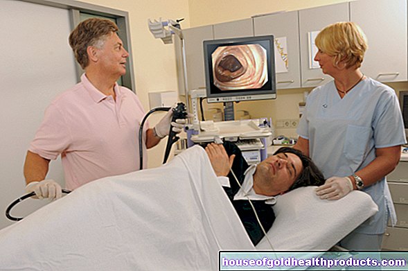
Cardiac catheter examination
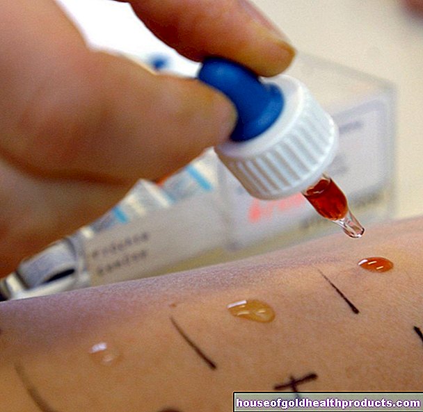
During a cardiac catheter examination of the left heart, a thin plastic tube (catheter) is usually inserted into an artery on the wrist or in the groin and pushed through the aorta to the aortic valve. Doctors use this test to identify coronary artery disease. This is especially important if a heart valve replacement is planned due to the aortic valve stenosis. Alternatively (and depending on the individual situation) doctors order a computed tomography of the heart with a contrast medium (cardio-CT).
Aortic stenosis: treatment
If the aortic valve stenosis is mild without symptoms, which is possible for about 50 percent of patients over a long period of time, conservative (non-invasive) treatment can be used first: the person affected should avoid physical exertion and take sufficient care of himself.
Moderate to severe aortic valve stenosis usually already causes symptoms. If patients with a high-grade aortic stenosis still have "no complaints", it is usually because they unconsciously take care of themselves physically so that no complaints occur. If additional symptoms are present in such patients (such as a pathological stress test, etc.) and in symptomatic patients, surgical therapy is recommended.
Aortic stenosis: TAVI and surgery
Doctors use different procedures for aortic stenosis:
Aortic valve replacement is particularly common for acquired stenoses. Doctors either operate on the open heart or insert a new valve in a minimally invasive manner as part of a cardiac catheter examination (TAVI = Transcatheter Aortic Valve Implantation). Younger patients with a low risk of surgery are usually operated on openly. Doctors also advocate surgery if additional interventions are necessary, such as a bypass.
The Ross operation is mainly performed in children with congenital aortic valve stenosis, but also in young and middle-aged adults. The aortic valve is replaced by the pulmonary valve, which sits between the right ventricle and the large pulmonary artery. This in turn is exchanged for a transplant. The procedure has many advantages (long valve life, good hemodynamics), but it is very complex and requires a donor valve.
If an operation cannot be performed, for example due to old age and concomitant diseases, doctors recommend a TAVI. As part of a cardiac catheter examination, they guide the new, still folded valve (usually a biological valve suspended from a metal mesh stent) on a catheter to the aortic valve. There a balloon pushes the metal grid apart, which finally anchors the valve between the chamber and aorta. To give the new valve space, the aortic valve stenosis is widened beforehand using a small balloon (balloon dilatation).
Balloon dilation alone (balloon valvuloplasty) is also used in children with congenital aortic valve stenosis. A valve replacement is problematic here because it cannot grow with the child. In the case of acquired aortic valve stenosis, balloon dilatation has a high rate of relapse (recurrence rate). Doctors therefore only use this method in an emergency in order to bridge the time until the final therapy.
Aortic stenosis: medication
Additional drug therapy aims to improve the symptoms until the operation. In particular, the consequences of aortic valve stenosis - heart failure and cardiac arrhythmias - are brought under control, for example, with beta blockers, cardiac glycosides or diuretics. A long-term effective treatment is not possible with medication, the stenosis must be removed in any case.
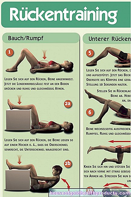
Exercise for aortic valve stenosis
There are no general recommendations for or against sporting activities in the case of aortic valve stenosis. The type and severity of the disease is always decisive.
Patients can find out whether sport is possible during the annual cardiological check-up. The attending physician examines the heart valve for possible damage and can make or update a recommendation for sporting activity.
Starting training for aortic valve stenosis
Before a patient with aortic valve stenosis starts exercising, an exercise ECG is necessary.
Aortic valve stenosis has long been considered an exclusion criterion for an exercise ECG. This also still applies to patients with symptomatic high-grade AS. However, especially in patients who show no symptoms, an exercise ECG can be helpful to reveal possible limitations in exercise capacity.
The stress ECG takes place under strict medical supervision, as undesirable side effects can quickly occur.
If a drop in blood pressure or cardiac arrhythmia occurs on the ergometer, the exercise must be stopped immediately.
After the examination, the cardiologist can use the data to assess the intensity with which the patient can be physically active.
Suitable exercise for aortic valve stenosis
Patients with asymptomatic valve stenosis can do sports of different intensity depending on the individual severity. Before the patient becomes physically active, their resilience should always be checked as part of a performance diagnosis.
The following overview shows which sport is possible with which degree of severity of the aortic valve stenosis:
Degree of severity light (no complaints, normal age-appropriate pump function in the heart echo, normal stress ECG):
Recommendation for physical activity: all types of sport are possible; including competitive sports
Degree of severity medium (normal pump function, normal stress ECG):
Recommendation for physical activity: Sports with low to moderate static and dynamic components: walking, cycling on the plain, golf, bowling, yoga, table tennis, volleyball, fencing, softball, archery, horse riding
Severity severe (impaired performance of the heart):
Recommendation for physical activity: no competitive sport; in individual cases for asymptomatic patients walking, cycling on the plain, golf, bowling, yoga
Always follow your doctor's recommendation if you have aortic valve stenosis. Before starting a new sport or changing the training plan, you should consult your doctor.
Aortic stenosis: disease course and prognosis
If aortic stenosis is left untreated, serious consequences can arise: For example, small blood clots can form as a result of turbulent blood flows in the calcified aortic valve. They can be swept away in the bloodstream and get into the brain. If they clog a vessel there and cut off the blood supply, it is called a stroke.
Aortic valve stenosis can also lead to abnormal heart rhythms. If left untreated, these can lead to ventricular fibrillation and cardiac death. Ultimately, progressive aortic valve stenosis causes increasing cardiac insufficiency which, without proper therapy, quickly ends in death.
However, with the right treatment for aortic stenosis, the prognosis is good.
Tags: first aid home remedies digital health