Femoral neck fracture
Dr. med. Mira Seidel is a freelance writer for the medical team.
More about the experts All content is checked by medical journalists.The femoral neck fracture (femoral neck fracture, femoral neck fracture) often occurs in older people with osteoporosis. The cause is usually a fall sideways on the hip or on the stretched or splayed leg. The hip fracture causes severe pain and the leg can no longer be actively moved. The therapy of choice is usually surgery. Find out more about the femoral neck fracture here.
ICD codes for this disease: ICD codes are internationally recognized codes for medical diagnoses. They can be found, for example, in doctor's letters or on certificates of incapacity for work. S72
Femoral neck fracture: description
The femoral neck fracture is a common fracture in the elderly. Young people are rarely affected as part of a high-speed trauma. Around 100,000 people in Germany currently suffer a femoral neck fracture every year. Women are about four times more likely to be affected than men. According to the Professional Association for Orthopedics and Trauma Surgery (BVOU), the lifetime risk of a femoral neck fracture is around 11 to 23 percent for women and five to 11 percent for men.
Forms of fracture: Classification according to Pauwels
The thigh bone (femur) consists of four parts: a long shaft, the knee joint, a short, slightly angled neck and the head, which together with the pelvis forms the hip joint. In the case of a femoral neck fracture, the part between the head and the shaft of the femur breaks. Depending on the course of the fracture gap, doctors differentiate between different types of fractures (according to Pauwels). This Pauwels classification is based on the size of the angle between the fracture surface and an imaginary horizontal line, and gives an indication of how stable the fracture is and which therapy option is to be considered:
- Pauwels grade I: fracture surface (indented) lies <30 degrees to the horizontal plane. There are no shear forces acting on the bone. This fracture of the femur can be treated conservatively.
- Pauwels Grade II: The angle (not indented) is 30 to 50 degrees. This type of fracture requires surgical treatment.
- Pauwels grade III: The angle (not indented) is greater than 50 degrees. It usually has to be operated on.
Femoral head necrosis
The femoral head is supplied from the front and back by blood vessels that radiate into the joint capsule. If these capsular vessels are torn off in a femoral neck fracture, there is a risk of femoral head necrosis (femoral head necrosis). The bony head of the thigh dies as a result of the reduced blood flow. The femoral head necrosis is one of the aseptic, i.e. not infection-related bone necrosis.
Femoral neck fracture: symptoms
If the hip is broken, severe compression and rotation pain in the groin and hip joint are the typical symptoms. This pain increases when the hip joint is moved passively (for example by the doctor). The patient can no longer actively move the leg. You may see local bruises and a bruise mark in the injury area.
If the femoral neck fracture is displaced, the leg appears shortened when the patient is lying down and is rotated outwards. Sometimes the thigh is also swollen in the area of the femoral head.
In rare cases, the symptoms of the compressed, firm femoral neck fracture can be very mild. Affected people often only go to the doctor after several days because of increasing pain under stress.
Femoral neck fracture: causes and risk factors
Due to the increase in the frequency of osteoporosis (bone loss) with age, the risk of a femoral neck fracture also increases with age. In the case of osteoporosis, due to the reduced bone density, little force is sufficient to break a bone. Even mundane falls, for example in the house or over a curb, can then end with a fracture of the thigh neck.
Some of the falls in the elderly are caused by dizziness or a brief loss of consciousness due to cardiovascular or nervous diseases. Medicines that restrict coordination and perception, alcohol and visual disturbances are also possible causes of accidents.
Very rarely, a femoral neck fracture occurs due to an existing tumor disease with daughter tumors (metastases) in the bone.
A femoral neck fracture is very rare in children, adolescents and adults under the age of 60. They need strong force on the bones in order for a fracture to occur, for example in an accident with a car or bicycle, in a skiing accident or a fall from a great height.
Femoral neck fracture: examinations and diagnosis
A femoral neck fracture can be life threatening. Therefore, if you suspect a femoral neck fracture, you should call the emergency medical service or your family doctor immediately. The specialist for all types of broken bones is the doctor for orthopedics and trauma surgery.
Medical history and physical examination
For the diagnosis, the doctor will ask you or your family members exactly how the accident happened and your medical history (anamnesis). Possible questions are:
- How did the accident come about?
- Can you remember the exact time of the accident?
- Was there any direct or indirect trauma?
- Where is the possible fracture?
- How do you describe the pain?
- Are there any previous illnesses such as osteoporosis, cardiac arrhythmias, diabetes or ametropia?
- Have you already had complaints such as stress-related pain?
Most of the time, the characteristic symptoms after a fall indicate a fracture of the femoral neck. The doctor will examine you carefully for more detailed clarification. He will also pay attention to injuries to the vascular and nervous system by checking your motor skills, sensitivity and blood circulation. He will also look for typical accompanying injuries to the skin and soft tissues.
Imaging procedures
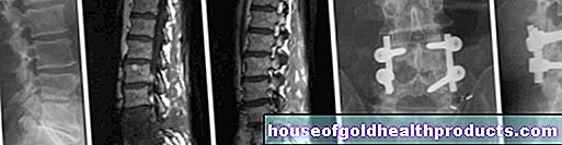
The diagnosis of femoral neck fracture is confirmed by x-rays of the hip in two planes. The X-ray image also shows exactly where the break is. This plays an important role for further therapy. Computed tomography (CT) is often done in order to plan the operation precisely.
If in advanced osteoporosis - despite clear symptoms - no break can be seen in the X-ray, further examinations are necessary. This includes a control X-ray three to five days after the accident and possibly a computed tomography (CT) or magnetic resonance tomography (magnetic resonance tomography, MRI) scan. If there is any doubt about the diagnosis of a femoral neck fracture, an MRI scan is the procedure of choice.
Femoral neck fracture - differential diagnoses
Other fractures in the femur or hip cause symptoms similar to those of the femoral neck fracture. These include, for example, the pertrochanteric femur fracture, the anterior pelvic ring fracture and the femoral head fracture (Pipkin fracture). The various fractures can be distinguished from one another with the help of X-ray diagnostics.
Femoral neck fracture: treatment
A femoral neck fracture is usually operated on. Only non-displaced, stable ("indented") fractures can sometimes be treated conservatively.
Older patients are often not very mobile after treatment of a femoral neck fracture - often for fear of further fractures - and therefore need help in everyday life. The aim of treatment is therefore that patients can put their leg back in place as quickly as possible. This is also important because if you lie down for long periods of time, muscle mass is rapidly reduced and complications such as pneumonia can occur.
Femoral neck fracture: conservative treatment
A wedged femoral neck fracture near the femoral head that is not displaced is called a stable femoral neck fracture. If the patient suffers only from minor pain, he does not necessarily have to operate, but can be treated conservatively. The injured leg is splinted and the patient is given pain medication and physiotherapy exercise.
Conservative treatment may also be indicated if a patient is not allowed to undergo surgery for certain reasons (e.g. if the general condition is risky).
Conservative therapy for a femoral neck fracture is unfortunately an exception, as the fracture is postponed in most cases.
Femoral neck fracture: OP
Different surgical methods are available for the femoral neck fracture, depending on where the fracture line runs. Basically, a distinction is made between femoral head preservation and femoral head replacement procedures. Which method is better in each individual case depends, among other things, on the age and condition of the patient, the type of fracture and the regenerative capacity of the bone tissue. Studies have shown that joint replacements often seem to be the better method for patients over 65 years of age.
The operation should be done as quickly as possible in the case of a femoral neck fracture: an operation within six to 24 hours after the trauma halves the risk of femoral head necrosis.
Femoral head conserving surgery
The femoral head conserving operation is more likely to be used in younger and active patients. The fragments are set back in the correct anatomical position and joined together with screws, plates and / or other implants. This procedure is called osteosynthesis. The prerequisite for this is sufficient blood circulation so that the bone can heal well.
Special screws such as the dynamic hip screw (DHS) or cannulated screw osteosynthesis are sometimes used. If the femoral neck fracture was only screwed or flattened, the leg must be consistently relieved for twelve weeks. With the other methods as well as with the dynamic hip screw, the patient is more mobile again.
Femoral head replacement surgery
In older patients, there is often little or no blood supply to the head of the femur. Then a joint replacement (endoprosthesis) is necessary: Either the femoral head (joint head) alone or the hip socket (joint socket) is replaced by an artificial prosthesis. In the latter case, one speaks of a total endoprosthesis (TEP, "artificial hip joint"). The advantage of this method is that the artificial joint can be loaded immediately, and the patient is usually quickly mobile again after the procedure. In the first few days after the operation, he can start with physiotherapy exercises.
A TEP is also used in patients with a fractured femur who also show wear and tear in the hip joint (coxarthrosis).
Femoral neck surgery: complications
Complications such as wound healing disorders, secondary bleeding, vascular or nerve injuries are relatively rare in a femoral neck fracture treated surgically. If the hip joint is replaced, more or less large blood losses are expected during the operation. There is a risk of blood clots (thrombosis) forming after the procedure.
Infections are a dreaded surgical complication of a femoral neck fracture. They can be tedious, but they are rare.
It is also rarely the case that the prosthesis shaft breaks out of the thigh bone. It then has to be replaced and fixed in another operation. This significantly delays the healing process.
In rare cases, a screw or plating operation worsens the blood flow to the femoral head. The femoral head can die off (femoral head necrosis) and must then be replaced by a prosthesis. The risk of this complication in connection with a femoral neck fracture increases with age, which is why an artificial joint is often used right from the start.
Further treatment
Before, during and for some time after the femoral neck surgery, patients receive thrombosis injections into the subcutaneous fat tissue to prevent blood clots (thrombosis) from forming. Wearing support stockings and doing physiotherapy can also prevent clots from forming.
Once the treatment of the femoral neck fracture has been completed, intensive physiotherapy should be started, which primarily trains the thigh muscles. The aim is for patients to be able to walk and climb stairs again as soon as possible.
Younger patients should partially load their leg for six weeks in a way that is adapted to the pain. Older patients, on the other hand, are advised to always fully load the leg in a way that is adapted to the pain. Pain-adapted means that the pain that occurs is always easy to bear.
Regular x-ray controls are important to check the position and consolidation of the fracture gap.
Femoral neck fracture: disease course and prognosis
If a femoral neck fracture is operated on soon, a very good result can usually be achieved. In individual cases, the prognosis can be estimated from the fact to what extent the femoral head has shifted and thus the blood supply is impaired. This Garden classification looks like this:
- Garden I: Such an indented abduction fracture usually has a good prognosis and a low necrosis rate.
- Garden II: This is an axially compressed adduction fracture that is not displaced. The risk of necrosis is low.
- Garden III: The adduction fracture is displaced without the posterior cortex being displaced. The necrosis rate is high.
- Garden IV: The fragments are completely displaced and the vascular supply is interrupted. There is a high rate of necrosis of the femoral head.
The steeper a fracture is, the more the shear forces increase under axial load, which shifts the fragments. This also increases the risk of femoral head necrosis and pseudoarthrosis (fracture that does not grow together).
Femoral neck fracture - healing time
The healing time for a femoral neck fracture varies from person to person. It depends on several factors, such as how old and active the patient is.
In patients under 65 years of age, with adequate treatment, the broken bone heals completely in up to 90 percent of cases. In the long term, however, femoral head necrosis occurs in almost 20 percent, which means that another operation with joint replacement is necessary.
According to the German Society for Trauma Surgery, patients spend an average of 11.7 days in hospital with the femoral head-preserving procedure, while a mean length of stay of 12.4 days can be expected for a joint replacement. With osteosynthesis, the implant is removed again after 12 months at the earliest.
Femoral neck fracture: prevention
There are simple steps you can take to prevent a fracture of the femur. If you suffer from another underlying disease such as cardiac arrhythmia, diabetes or ametropia, it is important to get this treatment. Because they increase the risk of falling and thus also the risk of a femoral neck fracture.
Sleeping pills are also dangerous for old people because they reduce the ability to react and thus also increase the risk of falling (e.g. when going to the toilet at night).
Hip protectors are only useful for patients who often fall, for example in nursing homes. These are specially developed underwear with pockets on the sides in the area of the hips. These bags contain plate-like, soft or hard protective elements made of various materials that can prevent hip fractures when falling.
A diet rich in calcium and regular physical activity make bones more stable so that they do not break easily. Supplement your diet with calcium- and vitamin D-containing supplements - your doctor will advise you in this regard. Measures such as age-appropriate furniture with handholds, walking aids and footwear adapted to the weather can also prevent a femoral neck fracture.
Tags: menopause teenager teeth


.jpg)