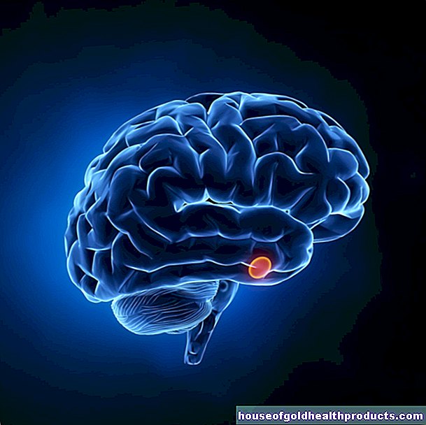
Astrocytoma
Ricarda Schwarz studied medicine in Würzburg, where she also completed her doctorate. After a wide range of tasks in practical medical training (PJ) in Flensburg, Hamburg and New Zealand, she is now working in neuroradiology and radiology at the Tübingen University Hospital.
More about the experts All content is checked by medical journalists.Astrocytoma is a brain tumor of the glioma type and can be benign or malignant. It is divided into pilocystic, diffuse and anaplastic astrocytoma and glioblastoma. The tumor can be operated on, irradiated, and treated with chemotherapy drugs. Depending on the severity, patients with an astrocytoma can be cured (grade I) or die after a short time (grade IV). Here you can read everything you need to know about astrocytoma.
ICD codes for this disease: ICD codes are internationally recognized codes for medical diagnoses. They can be found, for example, in doctor's letters or on certificates of incapacity for work. D43C71D33

Astrocytoma: description
The disease is assigned to the glioma as a brain tumor because it arises from the supporting tissue of the nervous system. With a share of over 60 percent, it is the most common glioma.
The World Health Organization (WHO) divides astrocytomas into four degrees of severity:
- Grade I: pilocystic astrocytoma
- Grade II: Diffuse astrocytoma
- Grade III: anaplastic astrocytoma
- Grade IV: glioblastoma
Both the treatment and the prognosis are based on this classification. Glioblastoma is the most common form of astrocytoma, with three new cases per 100,000 population per year. It is six times more common than diffuse and about ten times more common than anaplastic or pilocystic astrocytoma.
Pilocystic astrocytoma
This type of tumor is the most common brain tumor in children, but it also occurs in young adults. Mostly it grows in the area of the anterior visual pathway, the hypothalamus or in the cerebellum. It is well demarcated from healthy brain tissue and grows only slowly. This type of tumor is considered to be benign - it is extremely rare that it turns into a malignant tumor.
Diffuse astrocytoma
This type of tumor occurs mainly in adults between the ages of 30 and 40. It is usually found in the medullary bed of the cerebrum, the white matter, and grows irregularly into the neighboring tissue. However, it is only increasing slowly.
Diffuse astrocytoma is still considered benign to a limited extent, although it can almost never be cured. It can progress to the anaplastic type or glioblastoma over time.
Anaplastic astrocytoma
Anaplastic astrocytoma preferably affects adults between 35 and 45 years of age. It consists of degenerate (anaplastic) cells that usually grow relatively quickly. This tumor can either arise from the diffuse type or arise directly. Usually it degenerates further after a few months or years and turns into a glioblastoma.
Glioblastoma
Glioblastoma, the most common type of astrocytoma, can either arise from another astrocytoma - in which case the age peak of the patient is between 50 and 60 years of age. Or a primary “de novo” glioblastoma develops. This mainly affects older people in the sixth to seventh decade of life.
Glioblastoma
Further information on glioblastoma can be found in the article Glioblastoma.
Astrocytoma: symptoms
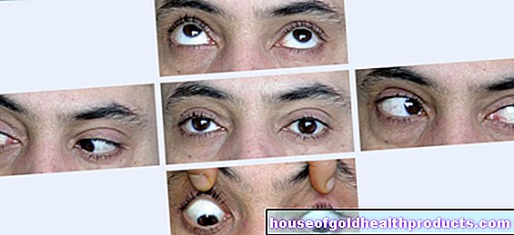
The symptoms that an astrocytoma causes depends on its exact location in the brain and its size. Pilocystic astrocytoma often grows along the visual pathway and causes visual disturbances. On the other hand, if it damages the thalamus, hormonal disorders can occur. If the tumor is in the cerebellum, it can lead to coordination problems, changed language and hand tremors.
Rapidly growing tumors not only displace individual brain structures, but also increase intracranial pressure. As a result, those affected can suffer from headaches, nausea and vomiting. In principle, an astrocytoma causes symptoms similar to other brain tumors.
Brain Cancer Symptoms
For more information on symptoms of a brain tumor, read the article Brain tumor - symptoms.
Astrocytoma: causes and risk factors
An astrocytoma is made up of so-called astrocytes. These cells form the majority of the supporting cells (glial cells) in the central nervous system. They demarcate the nerve tissue from the surface of the brain and the blood vessels. Just like other cells in the body, astrocytes are renewed regularly. Errors can occur that lead to uncontrolled cell growth and ultimately to a tumor.
Why an astrocytoma develops has not yet been adequately clarified. The only established risk factor is ionizing radiation. People are usually only exposed to harmful doses of radiation as part of radiation therapy.
In addition, this form of brain tumor occurs more frequently with certain underlying diseases. These include neurofibromatosis type I (von Recklinghausen's disease) and type II, tuberous sclerosis (Bournville-Pringle's disease), Li-Fraumeni syndrome and Turcot syndrome. These diseases are very rare and are usually associated with typical changes in the skin.
Astrocytoma: examinations and diagnosis
The right person to contact if an astrocytoma is suspected is the neurologist. He asks the person concerned precisely about their complaints, any previous and underlying illnesses as well as their living conditions (taking the medical history = anamnesis). He then initiates the further diagnostic steps and examinations.
The most important diagnostic procedure for an astrocytoma is magnetic resonance imaging (MRI) - an imaging procedure that uses magnetic fields and electromagnetic waves to generate precise cross-sectional images of the inside of the body. A contrast agent is often injected into a vein before the examination. Astrocytomas take up the contrast medium to different degrees. This creates a luminous area in the picture. A pilocystic astrocytoma lights up strongly and evenly, whereas a diffuse or anaplastic astrocytoma usually absorbs little or no contrast medium and appears rather dark. A glioblastoma, on the other hand, forms a characteristic ring-shaped structure with contrast medium.
If for various reasons an MRI cannot be performed, computed tomography (CT) is used as an alternative.
If you have a grade II or higher astrocytoma, a tissue sample should be taken and examined in the laboratory. In addition, further examinations may be indicated in individual cases, such as a collection and analysis of cerebral and spinal fluid (CSF diagnostics) or a measurement of electrical brain waves (EEG).
In principle, the same medical and technical examinations are carried out for an astrocytoma as for other brain tumors.
Astrocytoma: treatment
In general, this type of brain tumor can be operated on, stolen and treated with special drugs (chemotherapeutic agents). If a pilocystic astrocytoma is present, surgery is usually sufficient. Further treatment options are only used if the tumor has not been completely removed. A diffuse astrocytoma, on the other hand, can almost never be completely removed by surgery. If the remnants of the tumor that have remained start to grow again, they are usually irradiated.
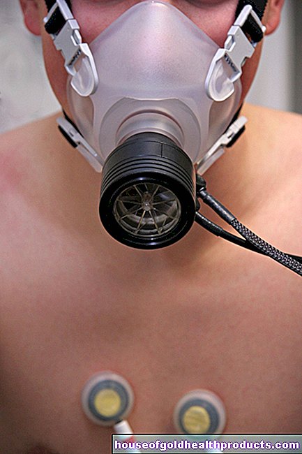
Brachytherapy can also be used for circumscribed tumors. For this purpose, small radioactive grains (seeds) are planted in the affected area of the body. The advantage of this form of radiation is that the tumor is hit very specifically and less healthy tissue is damaged.
If the astrocytoma enlarges even further under radiation, chemotherapy can be considered. Anaplastic astrocytoma is usually irradiated immediately after an operation or treated with chemotherapy drugs.
In addition to these therapeutic measures, there are also numerous treatment measures that do not fight the tumor itself, but the symptoms.Various medications can be administered for headaches, nausea and vomiting. In addition, those affected can take advantage of professional psychotherapy or pastoral care.
Pastoral tumor examination
You can find more detailed information on the examination and treatment in the article Brain Tumor.
Astrocytoma: disease course and prognosis
The course of the disease and prognosis differ between the individual forms of astrocytoma:
A pilocystic astrocytoma can be cured in the long term with complete surgical removal. If the tumor is in the vicinity of important brain structures such as the visual pathway, surgery is not always possible. The prognosis of alternative radiation or chemotherapy is somewhat worse. A total of 94 percent of all patients with this tumor type are still alive five years after diagnosis.
In contrast, an astrocytoma with WHO grade II or III has a significantly poorer prognosis. Second- and third-degree astrocytomas can usually be treated surgically, but often return (recurrences). Both can also become malignant and turn into a glioblastoma (grade IV). This has the worst prognosis: five years after the diagnosis, only five percent of those affected are still alive.
Thanks to good supportive therapeutic measures, the quality of life of patients with astrocytoma has improved.
Tags: interview home remedies nourishment