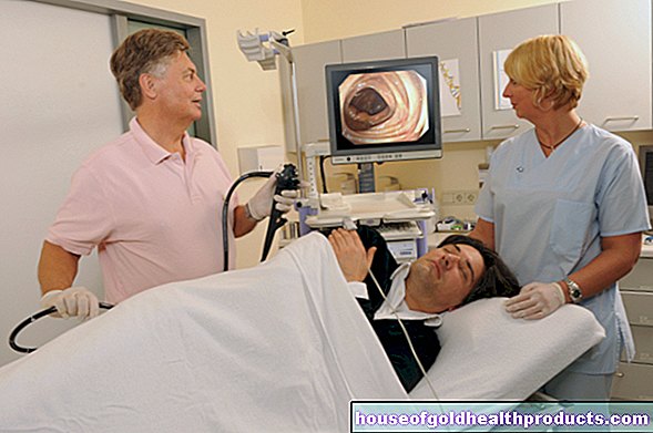
Otoscopy
Eva Rudolf-Müller is a freelance writer in the medical team. She studied human medicine and newspaper sciences and has repeatedly worked in both areas - as a doctor in the clinic, as a reviewer, and as a medical journalist for various specialist journals. She is currently working in online journalism, where a wide range of medicine is offered to everyone.
More about the experts All content is checked by medical journalists.During an otoscopy (ear or ear examination), the external auditory canal and the eardrum are examined, and if the eardrum is injured, the tympanic cavity is also examined. The ear, nose and throat doctor uses an otoscope - an instrument with an ear speculum, a light source and a magnifying glass. Find out everything you need to know about otoscopy here!

What is an otoscopy?
An otoscopy is a medical examination of the external ear canal and eardrum. The doctor usually uses an otoscope (ear mirror) - a medical instrument consisting of a lamp, a magnifying glass and an ear speculum. Sometimes an ear microscope is also used for the otroscopy, which offers a greater depth of field, brighter illumination and greater magnification. Then one speaks of ear microscopy.
When do you perform an otoscopy?
An otoscopy is one of the routine examinations at an ENT doctor. With their help you can, for example, detect foreign bodies in the ear canal and detect inflammation in the ear canal and on the eardrum, injuries, redness and bleeding. Bony growths (exostoses) that protrude into the ear canal can also be diagnosed in this way. If the doctor finds indentations or bulges in the eardrum during otoscopy, this may indicate an otitis media or fluid build-up behind the eardrum (tympanic effusion). Thickening of the eardrum and scars, on the other hand, are an indication of past inflammations or injuries.
In patients who tend to develop a lot of wax in the ears, otoscopy can be combined with regular cleaning of the ear canal.
In summary, an otoscopy is performed in the following cases:
- for the diagnosis and follow-up of various ear diseases such as otitis media
- if an injury to the ear canal or eardrum is suspected
- for the regular removal of ear wax
What do you do with an otoscopy?
No preparation for the otoscopy is required on the part of the patient. During the examination, the ENT doctor pulls the auricle slightly backwards and upwards, making the somewhat curved ear canal almost straight. After inserting the ear speculum, you can see the eardrum. During the examination, the patient should keep his head as still as possible in the position in which it was turned by the doctor in order to prevent the ear funnel from touching or even injuring the ear canal. If there is ear wax (cerumen), pus or flakes of skin in the ear canal, the ENT doctor will first remove these in order to have a clear view of the eardrum. In stubborn cases of wax, the ear is rinsed with lukewarm water - but only if it is certain that there is no damage to the eardrum.
Once the ear canal has been cleaned and the eardrum can also be seen, the ENT doctor can continue the examination and, if necessary, initiate treatment immediately. An example: In the case of otitis media with tympanic effusion, the doctor can make a small incision in the eardrum after anesthesia so that the effusion can drain out of the middle ear.
What are the risks of an otoscopy?
An otoscopy does not involve any risks or health hazards. However, it can be uncomfortable or even painful if there is inflammation in the area of the auricle, the ear canal, the eardrum or the middle ear.
What do I have to consider after an otoscopy?
There is nothing special to consider after a normal otoscopy. If the ENT doctor has also performed treatment, he or she can give special instructions such as not going to swimming pools or using certain medications (such as ear or nose drops for otitis media).
Tags: baby toddler prevention healthy feet