Chest x-ray
Updated onDr. med. Philipp Nicol is a freelance writer for the medical editorial team.
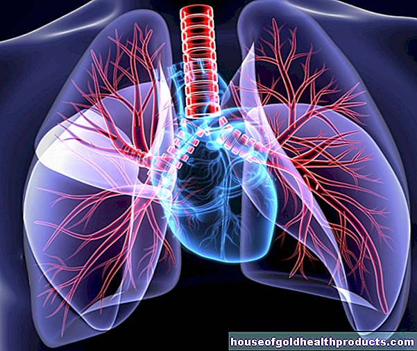
More about the experts All content is checked by medical journalists.A chest x-ray is a radiological examination of the chest and the organs in it, such as the lungs and heart. The examination can help diagnose a variety of diseases and is therefore often used. Read everything you need to know about the chest X-ray here.

What is a chest x-ray?
The chest X-ray is a standardized examination of the chest using X-rays. This examination is used to diagnose various diseases of the lungs, heart or blood vessels. Although computed tomography (CT) is becoming increasingly popular as an imaging method, the chest X-ray is still used frequently. One reason for this is the comparatively low radiation exposure (between 0.1 and 1 millisievert) - for comparison: With a computed tomography of the chest (CT thorax) it is 8 millisievert.
When is a chest X-ray performed?
A chest x-ray may be necessary for various complaints. Above all, these include:
- Chest pain
- Shortness of breath (dyspnoea)
- cough
- Difficulty swallowing (dysphagia)
- possible injury to the ribs
However, these complaints must not be isolated, but must always be assessed together with other factors such as the age of the patient, any underlying diseases and possible other complaints. It is also relevant whether the chest complaints are acute or have existed for a long time. Because of the radiation exposure, a so-called "justifying indication" must be available for each chest X-ray. This means that the value of the information from the X-ray image must outweigh the potential damage from the radiation. Only then will the lungs be x-rayed.
Basically, you should take a chest X-ray from the front (anterior-posterior) and from the side (lateral) so that you can assess the various structures well.
Every doctor should have a basic command of diagnosing a disease using a chest X-ray (diagnosis). However, the radiologists (X-ray specialists) specialize in this.
Chest X-ray: normal findings and findings typical of the disease
On a normal chest X-ray, the two lungs, the heart, the bony chest (including the ribs and collarbone) and the diaphragm can be assessed. The responsible doctor must pay particular attention to the following abnormalities:
Enlargement of the heart
A healthy heart should not be larger than half the chest diameter in the chest X-ray (heart-thorax quotient). Various heart diseases such as heart failure (heart failure) can lead to an enlargement of the heart, which can then be seen on the chest X-ray.
Fluid in the chest
As part of various diseases and injuries in the chest area (such as inflammation, heart failure, cancer or broken bones), fluid can accumulate in the so-called pleural space - the gap-shaped space between the pleura and pleura. Such a pleural effusion can be seen in the X-ray. Due to the force of gravity, the fluid collects at the lowest point of the chest, near the diaphragm.
The pulmonary edema must be distinguished from the pleural effusion. This is an accumulation of fluid within the lung tissue, often due to a heart disease. Pulmonary edema can also be diagnosed using a chest X-ray.
Pneumothorax
X-rays of the thorax usually allow a so-called pneumothorax to be reliably identified or ruled out. In the case of a pneumothorax, air has entered the pleural space and displaces the healthy lungs. An acutely life-threatening form is the tension pneumothorax, in which more air enters the pleural space with each breath and thus literally squeezes the lungs and heart. Both can be clearly seen on the chest X-ray.
Infiltrate
Lung infiltrate occurs when fluid and cells leak from blood and lymph vessels into the lung tissue. The reason is usually pneumonia. The lung infiltrate can be seen as a light-colored (compacted) structure on the X-ray.
What are the pros and cons of a chest x-ray?
The advantages of the chest X-ray are:
- available quickly and almost everywhere
- lower radiation exposure than with computed tomography
- high informative value for various diseases
The disadvantages of the chest X-ray are:
- Radiation exposure
- only one-dimensional images on which some structures "overlap" (with computed tomography, on the other hand, a spatial representation is possible)
Conclusion: The chest x-ray has become an indispensable part of medicine. If the radiation exposure is low, the examination quickly provides information about a wide variety of diseases and injuries in the chest cavity.
Tags: sleep interview toadstool poison plants