Urethral constriction
Clemens Gödel is a freelancer for the medical team.
More about the experts All content is checked by medical journalists.The urethral narrowing (urethral stricture) is usually based on a scarred change in the urethra. Men are particularly affected. The narrowing of the urethra is usually noticeable as a change in the urine stream or increased urinary tract infections. There are a number of surgical options for treating urethral strictures. Read more about symptoms, diagnosis and therapy of urethral constriction here!
ICD codes for this disease: ICD codes are internationally recognized codes for medical diagnoses. They can be found, for example, in doctor's letters or on certificates of incapacity for work. N35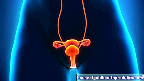
Urethral narrowing: description
The urethral narrowing (urethral stricture) is a common clinical picture in urological practice. Men are particularly affected: around one percent of them suffer from a narrowing of the urethra. Because of the shorter urethra, women have it much less often. A urethral narrowing can significantly reduce the quality of life and should therefore be treated early.
Urethral narrowing: symptoms
One of the main symptoms of a narrowing of the urethra is an altered urine stream. Usually the beam is weakened. However, it can also be changed in terms of its direction and shape (rotation, fanning). Due to the difficult urination, those affected often have to consciously press in order to urinate. This is not necessary if the urine flow is normal.
In addition, if the urethra is constricted, urination may be delayed when using the toilet, as the constriction has to be overcome first. A urethral stricture can cause urine to remain in the bladder after urination. This residual urine formation and the reduced urine stream increase the risk of urinary tract infections.
Those affected may also be worried by sudden interruptions in urination, "dripping" and uncontrolled loss of urine (incontinence). Another symptom of urethral constriction is the frequent need to urinate, but usually only small amounts of urine are excreted (pollakiuria). Blood in the urine (hematuria) and urinary stones are also common in urethral strictures.
Complication of urinary obstruction
In severe cases of urethral constriction, so-called urinary retention can occur, i.e. a complete blockage of the urethra. If this urinary retention persists, severe pain sets in and the urine can back up into the kidneys. Untreated kidney congestion leads to kidney failure - a life-threatening situation!
In men with a narrowing of the urethra, part of the erectile tissue of the penis (corpus spongiosum) can be affected by scarring. In the worst case, entire parts of the erectile tissue can scar. In this case one speaks of a cancellous fibrosis. The result is impaired erectile function of the penis.
Urethral narrowing: causes and risk factors
In around 30 percent of the cases, no explanation for the urethral narrowing can be found. Even in patients under 45 years of age, the cause of the urethral narrowing often remains unclear, or the stricture is the result of a pelvic fracture or a hypospadias operation. Hypospadias is a congenital malformation of the urethra: This is shortened and starts too early - in men, for example, on the underside of the penis, in women in the anterior vaginal vault.
In patients over 45 years of age, it is often medical interventions that lead to injuries and subsequent narrowing of the urethra.
In women, a narrowing of the urethra is usually due to a cramping (spasm) of the pelvic floor.
In men, urethral constriction often occurs in the anterior urethra, i.e. in the section between the pelvic floor and the penis. The posterior urethra, which is located between the bladder and the pelvic floor, is rarely narrowed. If a urethral constriction occurs here, the cause is usually a traumatic urethral tear or radiation therapy for cancer.
Causes in detail
The most common cause of urethral narrowing is injuries. This doesn't have to be major damage. Even microscopic injuries are sufficient for a scarred constriction, as can occur, for example, when placing a urinary catheter or during a cystoscopy. The majority of these interventions, however, have no negative consequences. Nevertheless, caution should be exercised in such invasive diagnostic and therapeutic procedures involving the urethra. With a frequent prostate operation, the transurethral prostate resection (TUR-P), up to five percent of the operated on later suffer from a urethral constriction. In women, incontinence operations in particular can lead to a urethral stricture. In addition, during childbirth, injuries to the urethra and subsequent narrowing of the urethra can occur.
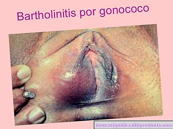
In around 20 percent of cases, (bacterial) inflammation of the urethra (urethritis) is the cause of the urethral narrowing. An important infection in this context is gonorrhea (gonorrhea), a sexually transmitted disease caused by bacteria of the Neisseria gonorrhoeae type.
Accidents can also lead to a narrowing of the urethra. This applies, for example, to pelvic fractures and blunt injuries in the step (“straddle trauma”), such as those caused by a bicycle fall. The urethra can be injured directly or due to the pelvic fracture and, in extreme cases, even tear off.
Congenital causes are responsible for five to ten percent of all cases of urethral narrowing. For example, some people are born with so-called urethral valves (sail-like membranes that constrict the urethra), a narrowing of the urethral opening (meatal stenosis) or malfunctions in the urethra (hypospadias).
Five percent of urethral strictures are caused by lichen sclerosus. This is an inflammatory skin disease that leads to hardening of connective tissue, especially on the glans of the penis and the foreskin.
There are also mechanical causes of urethral narrowing such as cancerous ulcers, polyps, pouches (diverticula), external pressure or a sinking of the pelvic organs (descent).
Urethral narrowing: examinations and diagnosis
The specialist in diseases of the urinary tract is the urologist. A narrowing of the urethra is suspected when patients report frequent urinary tract infections and changes in the urinary stream. Sometimes a urethral stricture is only noticeable through acute urinary retention.
To clarify the cause, the doctor will first take the medical history (anamnesis) and ask the patient the following questions, for example:
- What ailments do you suffer from?
- Have you noticed any changes in urination?
- Are you aware of any urinary tract disorders?
- Have you ever had any invasive urinary tract exams or treatments?
Then a urine test is done to rule out a urinary tract infection. This is important because otherwise, both diagnostic and therapeutic measures can lead to germs entering the bloodstream. The doctor calls this urosepsis ("blood poisoning").
The physical examination can identify externally visible changes, collect initial indications of a urethral stricture and perform an initial examination of the kidney.
The urologist can measure the urine flow with the so-called uroflowmeter. The patient has to urinate with a full bladder in a special toilet that measures the urine flow. If the urethra is narrowed, it takes longer to urinate and the urine stream is significantly weakened.
After this examination, ultrasound (sonography) can be used to determine whether there is any urine remaining in the bladder. The urethral constriction itself cannot usually be visualized with this procedure, but an assessment of the urinary bladder is possible. The layer of muscle in the wall of the urinary bladder may be thickened when the urethra is narrowed in an attempt to compensate for the increased resistance caused by the narrowing. The condition of the kidneys can also be assessed using ultrasound. Pay particular attention to evidence of urine backflow into the kidney.
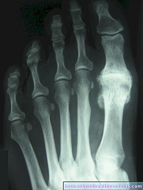
If these tests confirm a urethral constriction, the next step is to precisely determine its type, length and location. A so-called retrograde urethrography can be carried out for this purpose: the doctor injects a contrast medium at the exit of the urethra backwards into the urinary tract. Then an X-ray is taken. It enables conclusions to be drawn about the type of urethral constriction.
Alternatively, a similar x-ray examination with contrast agent - anterograde urethrography - can be performed. The contrast agent is injected either through a urethral catheter into the bladder or through a direct puncture of the bladder through the abdominal wall. The contrast medium can also be given into the vein, but then you have to wait until it reaches the urinary bladder. Urination can then be analyzed by an X-ray examination (micturition cystourethrography).
A urethraloscopy (urethroscopy) is mainly carried out if the urethrography has not provided any information about the narrowing of the urethra. The disadvantage of this examination, however, is that it does not provide any information about the length of the urethral constriction if the constriction cannot be overcome with the cystoscope.
So-called urodynamic examinations are carried out in special cases: With the help of measuring catheters in the rectum and bladder, the pressure conditions can be analyzed.
When diagnosing urethral constriction, benign and malignant tumors (such as the prostate) should be excluded as the cause of the symptoms. It is also possible that foreign objects (such as urinary stones) have entered the urethra causing a urethral stricture. Other causes such as megalourether, bladder neck sclerosis or detrusor bladder neck dyssynergy should be considered in unclear situations.
When examining a urethral narrowing, it is also determined whether and to what extent the erectile tissue is affected by the scarred change. This is important for therapy planning.
Urethral narrowing: treatment
Treatment of the urethral constriction must be planned individually. It depends on many factors, especially the length and location of the urethral constriction. But the amount of residual urine, possible kidney involvement and existing urinary tract infections also play a role.
As a rule, urethral constriction therapy consists of an invasive and sometimes difficult operation, which is best carried out in a specialized clinic. Various surgical techniques are available. None of them are fully suitable for all forms of urethral narrowing. To this day, experts have disagreed about the advantages and disadvantages as well as long-term results of the various techniques. It is therefore advisable to obtain a second opinion before starting therapy.
Dilation (bougienage)
Bougienage means stretching and is the oldest of all forms of urethral stricture therapy. In this treatment procedure, a special catheter is inserted into the urethra that can dilate the urethra (for example a balloon catheter). It is even possible for the patient to carry out the bougienage himself after a detailed explanation.
The most important problems with this procedure are, on the one hand, that the effect of a stretch only lasts for a certain period of time. As soon as the constriction occurs again, the stretch must be repeated. The first relapses can be expected four to six weeks after the bougienage. Over time, the intervals between the necessary applications usually become shorter.
On the other hand, the frequent insertion of the catheter can lead to minor injuries, which can worsen the urethral narrowing.
Bougienage should not be used in patients with acute urinary retention or severe residual urine formation. However, it is suitable for patients who refuse an operation or for whom the risk of anesthesia for an operation is too high.
Urethral slit
The urethral slit (urethrotomia interna) is usually only an option if the urethral narrowing is short (less than one centimeter) and the spongiofibrosis is only slight. In this case, the constriction can be split. To do this, the patient first receives general anesthesia or just spinal cord anesthesia. Then an endoscope is inserted into the urethra in order to split the scarred constriction in a controlled manner with a laser or a knife ("cold knife"). After the operation, a catheter should remain in place in the urethra for several days for splinting.
The cut in the scar creates a new wound, which in turn leads to scar formation. These scars are often larger than the originally treated scar and make the situation worse. Slitting a urethral constriction is therefore only successful in 50 percent of cases. It can be repeated, but this further increases the risk of relapse. The use of slitting should therefore be carefully considered.
Stent
With the help of an endoscope, a stent can be inserted at the site of the urethral constriction. A stent is a small tube made of a metal or plastic mesh that is designed to hold the urethra open. A distinction is made between permanent stents that can be left in place and temporary stents that have to be changed or removed after a few months.
As with bougienage, stent placement is accompanied by many possible complications. The stent can lead to recurring inflammation. It can also provoke new scarring. Overall, the long-term results of a stent for urethral narrowing are not good. This therapy method is therefore only used in exceptional cases.
reconstruction
With a recurring urethral constriction, open urethral surgery - urethral reconstruction - is usually performed. The narrowing of the urethra is cut out; the two ends of the urethra are tried to be sutured directly (end-to-end anastomosis). However, this is only possible with a short urethral constriction. If the indication is correct, the success rate is high.
If the urethral constriction is long (constrictions more than about four centimeters long), an operation with urethral replacement (urethral plastic) is usually performed. This procedure is also used for urethral ruptures. The foreskin and oral mucosa, but also other (mucous) skin areas of the patient, are used to reconstruct the missing section. The choice of urethral replacement depends on many factors. For example, studies have shown that the oral mucosa is in many cases quite suitable for reconstructing the urethra. However, after removal of the oral mucosa, various complications such as pain and sensory disturbances in the oral cavity can occur.
Urethral reconstruction is a very difficult procedure and should only be performed by an experienced surgeon. In the case of a complicated urethral narrowing, the operation can also be carried out in several sessions. There should usually be an interval of several months between each session.
After the operation, a catheter remains in the urethra as a splint for up to three weeks.
Overall, complications from urethral reconstruction are rare. Especially in young men, however, a urethra that has been shortened as a result of the operation can lead to erection problems. The result is that the penis curves downwards. During the operation it is also important to ensure that the erectile tissue is not disturbed in its function either directly or indirectly by blocking the blood supply or nerve damage.
Urethral narrowing: disease course and prognosis
An untreated urethral constriction can lead to a loss of kidney function and impairments in the quality of life due to urine congestion. For this reason, it is important that a narrowing is detected and treated early on.
However, after successful treatment, urethral narrowing may recur. Therapy for such a relapse is usually more difficult than initial therapy.
Overall, the following applies: the treatment results of a urethral constriction are the better, the closer the constriction is to the urinary bladder, the shorter it is and the less often the stricture has been treated.
Tags: womenshealth teenager drugs