Chorionic villus sampling
Nicole Wendler holds a PhD in biology in the field of oncology and immunology. As a medical editor, author and proofreader, she works for various publishers, for whom she presents complex and extensive medical issues in a simple, concise and logical manner.
More about the experts All content is checked by medical journalists.The chorionic villus sampling or chorionic biopsy is a voluntary examination from the field of prenatal (= prenatal) diagnostics. Using tissue from the placenta, chromosomal changes and genetic diseases can be detected in the unborn child at an early stage of pregnancy. Read more about the procedure, benefits and risks of the chorionic villus sampling here - and the differences to the amniotic fluid test.

Chorionic Villi Biopsy: What Are Chorionic Villi?
In the womb, the baby swims in the amniotic fluid in the amniotic sac. The outer membrane of this amniotic sac is the chorion, which forms the chorionic villi in early pregnancy. These finger-shaped protuberances form the contact point between maternal and child blood. This is where the exchange of oxygen and nutrients between mother and child takes place. From the 14th week of pregnancy (SSW), experts speak of placenta villi.
The villi are genetically derived from the fetus.Cells obtained from the chorion thus provide reliable information about hereditary diseases, congenital metabolic diseases and chromosomal disorders in the child.
Chorionic villus sampling: which diseases can be detected?
Chorionic villus sampling can identify diseases in the unborn that are due to an altered chromosome structure or number. Familial hereditary diseases (metabolic disorders) can also be identified. Among other things, the following can be verified:
- Trisomy 13 (Patau Syndrome)
- Trisomy 18 (Edwards Syndrome)
- Trisomy 21 (Down syndrome)
- Various hereditary metabolic diseases and other hereditary diseases such as cystic fibrosis, hemophilia (hemophilia) or muscle wasting (muscular dystrophy)
Malformations such as heart defects cannot be determined with a chorionic villus sampling. For this purpose, ultrasound examinations are necessary from about the 20th week of pregnancy. Neural tube defects such as an open back (spina bifida) or anencephaly (partial or complete absence of the cerebrum and other brain areas as well as the roof of the skull) can only be diagnosed with the amniocentesis. The same applies to malformations of the abdominal wall and blood group incompatibility between mother and child.
When is a chorionic villus sampling recommended?
If the risk of prenatally diagnosable diseases or chromosomal abnormalities is increased, your gynecologist will advise you to have a chorionic villus sampling. Such an increased risk exists in the following cases:
- The pregnant woman is older than 35 years.
- The pregnant woman has already given birth to a child with a hereditary disease or a chromosomal disorder.
- The pregnant woman or the father of the unborn child has a genetic defect.
- Hereditary diseases are known in the families of the expectant parents.
- Ultrasound examinations have found abnormalities in the unborn child (such as thickened neck folds).
When is the chorionic villus sampling performed?
A chorionic villus sampling is possible as early as the 10th to 12th week of pregnancy (SSW) and thus a little earlier than the amniotic fluid test (14th to 16th week of pregnancy).
How does the chorionic villus sampling work exactly?
After a detailed medical consultation about the benefits and risks of the chorionic villus sampling, you must agree to the procedure in writing. The entire examination is carried out on an outpatient basis and usually without anesthesia. First, your gynecologist will check the position of the placenta as well as the position and age of the child in an ultrasound examination. This is followed by the actual chorionic villus sampling, whereby there are two possible procedures: the so-called transcervical (via the vagina) or the transabdominal chorionic villus sampling (through the abdominal wall). With both procedures, the fruit cavity remains untouched, so that injury to the child is unlikely.
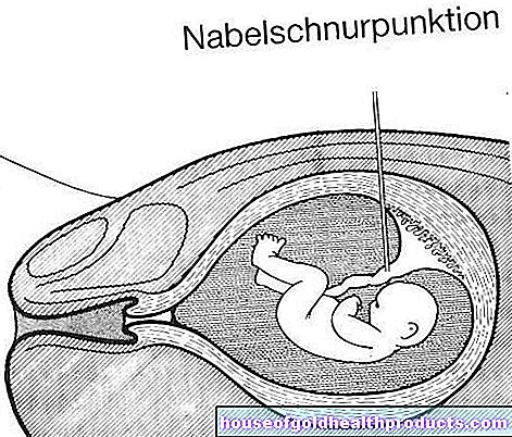
Transabdominal chorionic villus sampling: In an ultrasound examination, the doctor first selects a suitable puncture site. There he then inserts a thin puncture needle over the abdominal wall and carefully pushes it into the placenta in order to remove a small amount of tissue (20 to 30 milligrams) from the chorion. The doctor carefully controls the entire process using the ultrasound monitor.
Transcervical chorionic villus sampling: In the transcervical chorionic villus sampling, tissue is not removed with a needle through the abdominal wall, but with a thin catheter through the vagina and the cervix. The thin tube is carefully pushed up to the placenta under ultrasound control in order to obtain tissue from the chorionic villi.
The child's chromosomes are then extracted from the tissue samples in the laboratory and examined more closely. If necessary, a cell culture is set up for a DNA analysis.
After the chorionic villus sampling
Most pregnant women find the procedure itself uncomfortable, but not very painful (similar to taking a blood sample). Afterwards, some women complain of a kind of cramping or a feeling of pressure in the abdominal area, which, however, subsides after a few hours.
You can leave the hospital or practice about half an hour after the chorionic villus sampling. However, you should take it easy for the next three days and avoid sexual intercourse. Your gynecologist will examine you again in the following days. If you experience pain, discomfort and bleeding or if you lose amniotic fluid, you should consult a doctor immediately!
When are the results of the chorionic villus sampling available?
A major advantage of the chorionic villus sampling is that the finding is available within a few days at best. If, for example, a serious hereditary disease is found in the child and the pregnant woman then decides to terminate, this can be done in the first trimester. At this point, an abortion is physically and mentally easier for women to cope with than in the second trimester.
The results of the chorionic villus sampling are 99 percent certain. However, misdiagnoses and inconclusive findings are possible. If the removed cells do not show a uniform picture (mosaic findings) or if maternal cells were accidentally punctured, follow-up examinations may be necessary. If a cell culture has to be set up for further examinations if the result is unclear, it can take up to two weeks to obtain a final result.
How safe is the chorionic villus sampling?
Every intervention involves dangers. The risk of miscarriage is greater with chorionic villus sampling (around one percent) than with amniocentesis (0.5 percent). This is mainly due to the fact that the natural miscarriage rate is generally higher in early pregnancy than in the weeks after. Other risks are:
- Infections
- Vascular injuries
- premature labor
Successful chorionic villus sampling requires experience and skill. Therefore, have the procedure carried out in a specialized center or by an experienced doctor.
Chorionic villus sampling: what to consider
Before any prenatal diagnosis (examination of the unborn child in the womb), you should consider what consequences a positive result would have for you personally. Chorionic villus sampling is a fairly reliable method of identifying abnormal chromosomes or hereditary diseases in unborn babies. However, it is impossible to predict how severely the child will be impaired. In addition, the chorionic villus sampling does not guarantee a healthy child!
Tags: smoking Diseases symptoms