Ventilation
Valeria Dahm is a freelance writer in the medical department. She studied medicine at the Technical University of Munich. It is particularly important to her to give the curious reader an insight into the exciting subject area of medicine and at the same time to maintain the content.
More about the experts All content is checked by medical journalists.With ventilation, the patient's breathing is supported or replaced. It is always used when the person concerned cannot breathe himself or only inadequately. A distinction is made between various ventilation techniques. Read everything about ventilation, how it works and the risks it carries.

What is ventilation?
Ventilation replaces or supports the breathing of patients whose spontaneous breathing has failed (apnea) or is no longer sufficient to maintain bodily functions. The carbon dioxide content in the body increases while the oxygen content falls. The effectiveness can be measured by blood gas analyzes, by measuring the absorption of light when x-raying the skin (pulse oximetry) or the concentration of carbon dioxide in the exhaled air (capnometry). A basic distinction is made between non-invasive ventilation (NIV ventilation) via a mask and invasive ventilation (IV) via a tube in the windpipe (tube). Depending on the clinical picture, different forms of ventilation are used.
Controlled ventilation (continuous mandatory ventilation; CMV ventilation)
The ventilation machine (respirator) takes over the entire work of breathing, regardless of whether the patient is breathing naturally.
Assisted spontaneous breathing (ASB ventilation)
With assisted ventilation or assisted spontaneous ventilation, the greater part of the work of breathing and breathing regulation is done by the patient himself. The ventilator provides support like an additional respiratory muscle. One also speaks of machine-assisted spontaneous breathing.
When is ventilation performed?
Ventilation is always necessary when natural spontaneous breathing is insufficient to inhale enough oxygen and exhale carbon dioxide. Depending on the cause, the doctor selects the appropriate ventilation modes.
In the case of chronic obstructive pulmonary diseases (COPD) or diseases with weak respiratory muscles, ventilation during the night is sufficient to allow the respiratory muscles to recover. This can also be carried out as home ventilation with respirators at home. Lung failure, caused for example by poisoning, pneumonia or embolism, can make short-term ventilation necessary. Ventilation is also necessary during anesthesia, as the anesthetics switch off spontaneous breathing. If patients can no longer breathe independently due to paralysis or a coma, long-term mechanical ventilation ensures the supply of oxygen.
What do you do with ventilation?
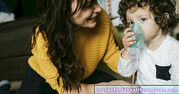
In contrast to spontaneous breathing, artificial ventilation presses air into the lungs using excess pressure. The non-invasive artificial ventilation uses masks that are put over the mouth and nose, while the invasive ventilation takes place via a tube that is pushed through the mouth or nose into the windpipe (intubation).
Controlled ventilation
With so-called controlled ventilation, the respirator takes over the entire work of breathing and is not influenced by the patient's inhalation and exhalation.
If a certain volume is to be given per breath in a given time, volume-controlled ventilation (VCV) is used. When the appropriate tidal volume is reached, exhalation is initiated. VCV ventilation can also be distinguished once again as to whether the pressure in the lungs remains high during exhalation (continuous positive pressure ventilation, CPPV) or decreases again (intermittent positive pressure ventilation, IPPV ventilation). .
For pressure-controlled ventilation (PCV ventilation), the ventilator creates a certain pressure in the airways and the alveoli so that as much oxygen as possible can be absorbed. As soon as the pressure is high enough, exhalation starts.
Assisted ventilation
Here the respirator only starts ventilation when the patient inhales himself. The breath is the starting signal (trigger) for the support by the ventilation machine (assist-control ventilation, A / C). A distinction is made as to whether the trigger for the respirator should be measured via the inhaled volume (volume-support ventilation, VSV ventilation) or the pressure (pressure-support ventilation, PSV ventilation).
Synchronized intermittent mandatory ventilation (SIMV ventilation)
With SIMV ventilation, assisted spontaneous breathing by the patient is combined with controlled ventilation. The respirator supports the patient when the patient is forced to breathe. The interval between two inhalation phases is determined. If the patient now breathes outside of these distances, he breathes independently without assistance. If the patient does not breathe freely, the respirator ventilates independently.
High-frequency-oscillation-ventilation; HFO-ventilation
High-frequency ventilation has a special position and is mainly used in children and newborns. HFO ventilation creates turbulence in the airways so that the air in the lungs is constantly mixed. This leads to an improved gas exchange despite the low ventilation volume.
CPAP ventilation
With CPAP ventilation, the patient's spontaneous breathing is supported by permanent positive pressure (PEEP). The patient determines the depth and frequency of breathing himself, but can breathe in more easily and draw more oxygen from the air because the alveoli are kept open by the excess pressure. This form is often used as therapy for respiratory arrest during sleep (sleep apnea). PEEP (positive end-expiratory pressure) describes the pressure that remains in the lungs at the end of exhalation. If this pressure is too low, the alveoli cannot be kept open and they close (collapse).
What are the risks of ventilation?
In addition to skin irritation or wounds from the mask or tube, there may be complications from the ventilation itself. These include:
- Damage to the lungs from pressure
- lung infection
- Increase in pressure in the chest
- Abdominal distension
- Decrease in venous return to the heart
- Increase in vascular resistance in the lungs
- Decrease in the heart's pumping capacity
- Reduced blood flow to the kidneys and liver
- Increase in intracranial pressure
Lung-protective ventilation reduces or prevents such damage by limiting ventilation pressures and ventilation volumes.
What do I have to consider after ventilation?
While short-term ventilation rarely causes problems, long-term ventilation can lead to habituation effects. The own respiratory drive decreases and the auxiliary respiratory muscles are reduced. In order for the patient to learn to breathe independently again, mechanical ventilation is gradually reduced in a weaning phase. Your own breath drive is encouraged and your muscles are rebuilt bit by bit.
Tags: menopause organ systems elderly care