Lithotripsy
All content is checked by medical journalists.Lithotripsy is the disintegration of gall, urinary and kidney stones. These collect in the ducts of the organs and cause severe pain. Various treatment methods are available for the removal of bile, urine and kidney stones. Read all about lithotripsy procedures, when they are performed and what the risks are.

What is lithotripsy?
Doctors understand lithotripsy to be the breaking up of so-called concretions, i.e. deposits such as kidney, urinary or gallstones that arise from various substances. Lithotripsy may be necessary if these symptoms are the cause. There are various methods to choose from:
- Extracorporeal shock wave lithotripsy
- Ureteroscopy
- Percutaneous nephrolitholapaxy
- Bile duct specimen
When do you perform lithotripsy?
Lithotripsy can be necessary if stones (concretions) form within organs that the body can no longer break down or excrete on its own. For kidney stones, for example, doctors recommend lithotripsy from a diameter of five millimeters. With smaller stones, conservative measures such as drinking plenty of fluids, applying heat or physical activity are usually sufficient.
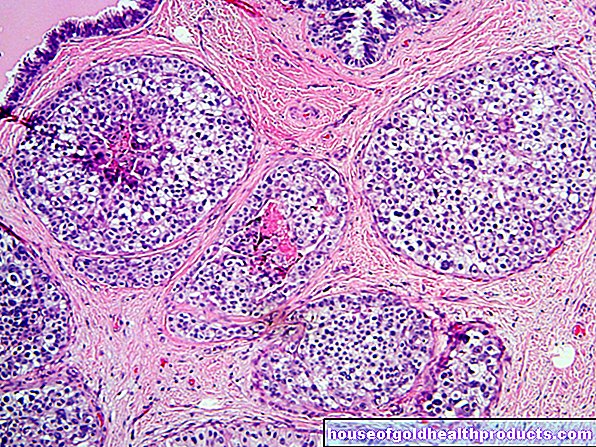
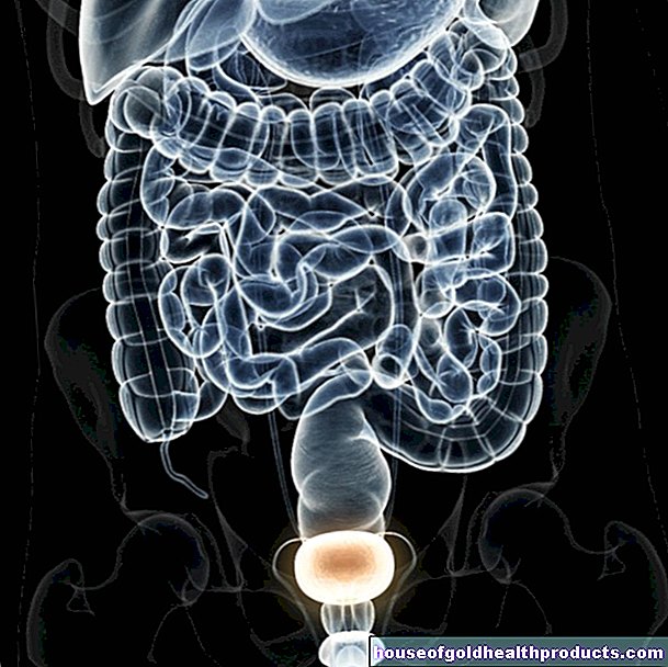
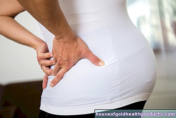
Kidney and urinary stones
The kidneys are responsible for producing urine. They filter the blood and excrete toxins, salts and excess water, among other things. The urine then flows through the ureters into the bladder, where it is stored.
Urine is made up of various substances that can accumulate and form stones in the event of certain illnesses, low levels of drinking or poor nutrition. These are initially only found in the kidneys, later they dissolve and are flushed into the bladder with the urine. Small stones cannot be felt, but large ones get stuck in the ureter and cause severe cramp-like pain (colic) there. There is also the risk of kidney failure because the urine backs up into the kidneys and damages them.
Gallstones
Among other things, the liver produces bile, which plays an important role in fat digestion and is stored in the gallbladder.
In many older people, the gallbladder contains stones, but these usually do not cause any symptoms. If a gallstone loosens and enters the bile duct, severe, colicky pain, nausea, and vomiting can occur, similar to kidney stones.
In the event of complaints, bile duct stones are usually surgically removed endoscopically; in the case of stones in the gallbladder, gallbladder removal as part of a laparoscopy is an option. Lithotripsy is used less often.
What do you do with a lithotripsy?
Pain reliever drugs not only reduce the patient's stress, but also relax the muscles of the excretory ducts. This facilitates the elimination of the stone that has been shattered after the lithotripsy.
Extracorporeal shock wave lithotripsy
Extracorporeal shock wave lithotripsy (ESWL) is a leading procedure in the removal of urine and kidney stones. Shock waves - particularly strong sound waves - that are generated with the help of a device outside the body (extracorporeal) are aimed directly at the stone. This breaks into small pieces and can then be removed endoscopically or excreted.
Ureteroscopy
With a ureteroscopy, the doctor inserts a semi-rigid or flexible instrument into the ureter through the urethra and bladder. Once he has reached the stone, he performs the lithotripsy with a laser attached to the instrument that generates the shock waves. The individual fragments are removed with special gripping instruments or later excreted with the urine. The ureteroscopy is used, for example, after an unsuccessful shock wave lithotripsy.
Percutaneous nephrolitholapaxy
Percutaneous nephrolitholapaxy (PCNL) is an endoscopic, i.e. surgical, kidney stone removal under general anesthesia, in which only a small incision is necessary. The doctor first inserts a special splint into the ureter of the affected side through which the urine flows. During the operation, the kidney is checked with an ultrasound machine or with the help of a computer tomograph.Now he sticks with a hollow needle (cannula) through the skin (percutaneously) in the direction of the kidney and then introduces a nephroscope, i.e. a device for kidney mirroring, through the puncture canal. The actual lithotripsy is carried out with ultrasound or laser probes. The stone fragments are flushed out through the ureter and the wound canal sutured.
Bile duct specimen
In the case of a bile duct specimen, the doctor inserts a medical instrument (endoscope), with which he performs the lithotripsy in the bile duct, through the mouth, esophagus and stomach into the small intestine. With the help of an X-ray machine, he checks the common duct of the gallbladder and the pancreas, which opens into the small intestine. In order to prevent a new blockage of the bile duct, the surgeon often widened the opening of the excretory duct in the small intestine with an incision. The stone fragments are usually excreted through the intestines with the help of the normal flow of bile.
What are the risks of lithotripsy?
Extracorporeal shock wave lithotripsy can cause pain that is treated with painkillers. In addition to bleeding, cardiac arrhythmias may occur as a possible consequence of the treatment.
There may be blood in the urine for a short time after the ureteroscopy, occasionally the patients have a fever and are prescribed antibiotics. In very rare cases, the ureter is injured - up to and including a complete tear - during the procedure. Very rarely does the ureter scar after lithotripsy so much that the urine can no longer flow into the bladder and backs up into the kidney.
With a bile duct specimen, the endoscope can injure or puncture the intestinal wall or bile ducts. Larger injuries have to be operated on again. Bleeding, infections of the bile ducts or acute pancreatitis are also possible after lithotripsy as part of a bile duct specimen.
If the cause of the stone formation persists, stones can reappear after lithotripsy and require renewed or extended therapy.
What do I have to consider after a lithotripsy?
After a lithotripsy, watch out for warning signs to quickly spot complications. These include, for example, pain, discomfort or blood in the urine. After removing gallstones as part of a bile duct specimen, you should also look out for red or black stools and diarrhea. In these cases, contact your doctor immediately.
In order to avoid new stone formation with another lithotripsy, the treatment of a possible underlying disease and an appropriate diet are particularly helpful. These include, for example, a low-salt and low-protein diet and adequate fluid intake.
Tags: Diagnosis drugs organ systems

.jpg)