Heel foot
All content is checked by medical journalists.A pes calcaneus is a noticeable deformity of the foot that occurs mainly in babies and is mostly caused by its position in the womb. In some cases it is also a symptom of another disease. In most cases, a hook foot can be treated well. Read here which therapy the doctor recommends!
ICD codes for this disease: ICD codes are internationally recognized codes for medical diagnoses. They can be found, for example, in doctor's letters or on certificates of incapacity for work. Q66M95
Brief overview
- What is a hook foot? The malalignment of the feet is usually congenital, but sometimes also arises from illnesses or accidents. The foot is strongly bent upwards, in extreme cases the toes lie against the shin.
- Treatment: For newborns mostly spontaneous healing, physiotherapy, plaster and splints, surgery, special shoes
- Causes: Baby's constrained position in the womb, viral infections, genetic causes, neurological diseases, accidents
- Diagnosis: assessment of the visible symptoms, imaging tests, gait analysis
- Prevention: In the common primary form, careful treatment of previous illnesses and injuries is not possible
What is a hook foot?
A pes calcaneus is a special misalignment of the foot. It is either congenital or acquired during life. This secondary hook foot is the result of another disease. In most cases, babies are born with one hook foot. Less common is a combination of crooked foot and crooked foot, which is known as heel-toe-toe or toe-toe-toe (pes valgocalcaneus).
It is estimated that almost every second newborn is affected by a hook foot. The deformity is differently pronounced. In the best case scenario, it is barely recognizable. In most cases, the congenital carpus heals completely. With the secondary variant, the prognosis depends on the underlying disease.
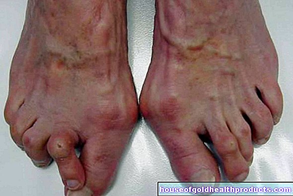
Symptoms: This is what a hoe foot looks like
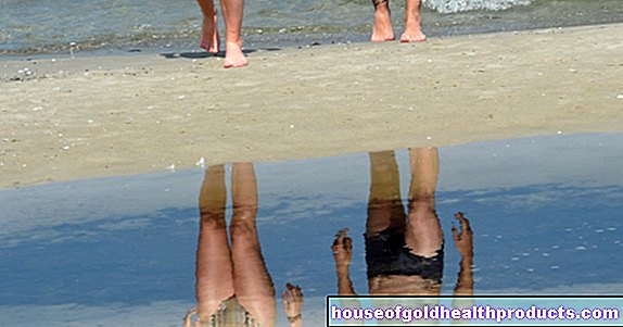
A pronounced heel foot is noticeable. The entire foot is stretched up towards the shin. Medical professionals refer to this symptom as dorsiflexion. This hyperextension means that it is not possible to bend the foot normally downwards (plantar flexion). In extreme cases, the toes lie against the shin so that the sole of the foot points outwards. The foot looks like it has been folded. Visually, it is the opposite of the equinus, in which the toes point down.
As a rule, the deformations only affect the soft tissues, the bones are not affected. Therefore, this deformity can usually be treated well. A congenital hook foot with deformed bones, on the other hand, is very rare.
With the buckle-heel foot, the sole of the foot is also hyperextended upwards towards the shin. In addition, the ankle is slightly bent inwards, which is why the sole rotates a little outwards.
Possible complaints from consequential damage
It is not possible to walk normally if the heel foot is noticeable. Even if the deformity is less pronounced, it should definitely be treated - if it does not resolve on its own. This is important to avoid consequential damage. Because even a slight heel foot affects the entire musculoskeletal system.
The gait pattern changes, the leg muscles and tendons are overstretched. This particularly affects the calves. The load on the heel is too great, which is why those affected often get pressure points. All of this makes movement difficult and causes pain. Overall, an imbalance develops between muscles and joints. Many patients instinctively take a relieving posture to relieve the painful areas. This brings other health problems with it, for example in the back.
How is a hook foot treated?
Treatment of the foot pusher primarily depends on the cause. A hoe foot in babies usually heals even without therapy.
Spontaneous healing
The hoe foot in babies is a common foot misalignment. However, doctors do not have to treat it automatically because in many cases it will resolve on its own. This sometimes happens within a few days after the birth.
Massage and physiotherapy
If the malalignment of the foot does not normalize shortly after birth, doctors treat the hoof foot in babies. Manual mobilization comes first: muscles and ligaments are massaged and stretched until the sole of the foot has returned to its normal position.
It is recommended that parents support this process by having the physical therapist show them exercises that they can do at home with their child. If necessary, as the children get older, they can do exercises themselves under supervision. But that is seldom necessary.
Treatment with plaster of paris or splints
Therapy for the baby's hook foot is usually complemented by a treatment called redression. Put simply, it is about forcing the foot into the correct posture and fixing it there until the structures have adapted and the foot remains in this position. This takes place in two phases.
In the first step, doctors carefully press the foot into the normal position and hold it there with special bandages or a cast. In the next step, the child receives specially adapted splints for the night. They fix the foot in an overstretched position, which actually corresponds to the equinus. In this way, the doctors ensure that the joint has the necessary mobility.
surgery
Surgery is very seldom necessary for the baby's congenital hook foot. Doctors use them more often in the secondary form. If the deformity cannot be eliminated by conservative measures, there are various options for treating it surgically. There are various methods available to surgeons for this purpose. These are the most important:
- The Achilles tendon connects the calf muscle to the heel bone. In the case of a hoe foot, it is permanently overstretched. It is therefore advisable to shorten it or change its position in order to exert tension on the sole of the foot.
- It has a similar effect when surgeons insert additional muscle tendons in the area of the Achilles tendon to strengthen them and thus increase muscle tension.
- In some cases, surgeons remove a piece of bone from the heel bone (a hindfoot osteotomy) to help the foot return to normal position.
- Another possibility is to force the foot into the correct position and fix it there permanently. Doctors stiffen the ankle joint, for example with a screw (arthrorise). However, this variant limits the patient's mobility in the long term. This can be felt, for example, when walking quickly or when running.
Insoles and special shoes
A hoe foot that arises as a side effect of a disease is not always completely curable. Treatment therefore includes wearing insoles or special shoes, for example, in order to improve movement sequences and avoid further consequences for the musculoskeletal system.
How is a heel foot made?
When considering the possible causes of a pes calcaneus, it is important to distinguish between the congenital and the acquired variants.
Congenital hook foot
The congenital hook foot in babies is either a disease in its own right or it occurs as a result of another disease. Accordingly, there are various causes.
The common primary tarpaulin, which in most cases heals without any problems, is probably caused by the position of the child in the womb. If pressure is exerted on the baby's feet due to a lack of space, they initially remain in the corresponding misalignment. A spontaneous resolution within a few days is likely.
There are also genetic causes. Some children have an imbalance in the muscles between the lower legs and feet. The calf muscles are then relatively weak, which is why the muscles in the area of the shin and the back of the foot pull the foot upwards.
A disease of the nerves (neurological disease) can also lead to a hoe foot in the baby if it interferes with the control of the corresponding muscles. A typical example is a so-called open back (spina bifida), i.e. a malformation of the spine and spinal cord, which may lead to symptoms of paralysis. The same goes for brain damage. As a rule, it also reduces the response to the muscles by the nerves.
Acquired hook foot
The secondary hook foot occurs in principle at any age. Possible causes are inflammation, such as those caused by the viral disease poliomyelitis (polio). In many cases they lead to paralysis and thus also to a hoe foot. Thanks to extensive vaccinations in Germany, polio is considered to be eradicated. The autoimmune disease myasthenia gravis, for example, has a similar effect.
A hook foot can also develop if the Achilles tendon is injured or possibly severed. It helps keep the foot in place. The calf muscles also play an important role. If they are injured or no longer properly cared for because the corresponding nerve is damaged in an accident, for example, this often leads to an imbalance in the muscles and, as a result, to misalignments of the foot.
Surgery is also a possible cause of tarnish foot. This is especially true if the doctors want to correct another foot misalignment and the correction is too intensive, for example due to an excessive lengthening of the Achilles tendon. Even a permanently incorrectly positioned foot can lead to misalignments.
How is a hook foot determined?
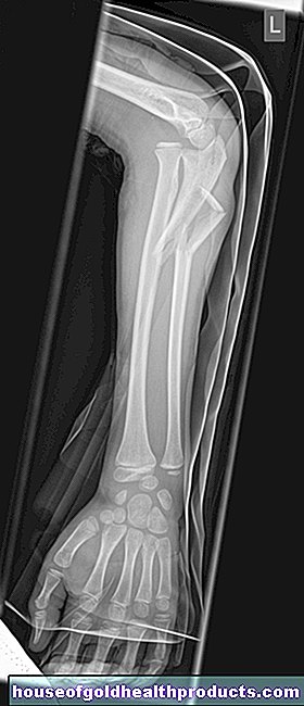
The doctor usually recognizes a hook foot in a baby by the visible symptoms, i.e. the upturned toes. If necessary, he will conduct additional examinations to determine the extent of the foot malposition. Among other things, he clarifies the question of whether only the soft tissues or also the bones are deformed.
In the case of a newborn, a comprehensive diagnosis is particularly relevant if the hook foot has not regressed after a few days. Some of the examinations are also important in order to identify or rule out other diseases as the cause.
In a conversation with the parents or the adult affected, the doctor clarifies which relevant previous illnesses exist (anamnesis). With a neurological examination, he checks the function of the nerves and looks for disorders or failures such as symptoms of paralysis.
Imaging methods such as x-rays, ultrasound, magnetic resonance imaging (MRI), and computed tomography (CT) help determine the exact extent of the phalanx. A gait analysis is useful in older patients.
If the disease has progressed, further examinations may be required. The doctor assesses the extent to which consequential damage has already occurred to the musculoskeletal system. The focus is on the knees, pelvis and spine.
Prevent
It is not possible to prevent primary tarnish in babies. If diseases are diagnosed during pregnancy that may lead to a secondary hoe foot, such as an open back, these are treated comprehensively.
With the acne heel that has been acquired, it is possible, at least to a small extent, to prevent the malalignment of the foot. For this it is advisable to do sport regularly and then to stretch muscles and ligaments. This ensures a better balance of the muscles and maintains mobility.
After injuries, it is important to fix the foot in the correct position for the healing process in order to prevent a heel foot.
Tags: vaccinations healthy workplace digital health