Thoracoscopy
Updated on All content is checked by medical journalists.Thoracoscopy is an endoscopic procedure used to examine the pleural cavity. This is the gap-shaped space between the two sheets of the pleura. Thoracoscopy is used, for example, if lung cancer is suspected or pleural effusions of an unclear cause. The doctor can also perform small interventions during this procedure, such as taking tissue samples. Read everything you need to know about thoracoscopy here.

What is a thoracoscopy?
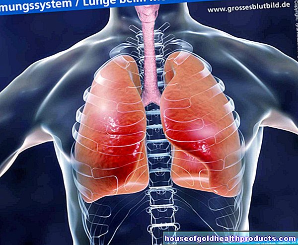
Thoracoscopy is an examination method from the field of pulmonary medicine. With their help, the doctor can examine the chest cavity more closely: To do this, he introduces endoscopic instruments, including a camera, a lamp, irrigation and suction devices, into the pleural space - the gap-shaped space between the pleura and pleura. The method is comparatively simple and is very informative. It can be carried out either on an outpatient basis or as part of an inpatient stay.
Nowadays the procedure is mostly performed as video-assisted thoracoscopy (VAT). The doctor can also perform minimally invasive interventions during the examination, for example taking a tissue sample from the pleura or removing a lung flap (in the case of lung cancer). Doctors then speak of video-assisted thoracoscopic surgery (VATS).
When is a thoracoscopy performed?
The pleural cavity is examined by means of thoracoscopy, i.e. the gap-shaped space between the inner and outer sheets of the pleura (pleura and pleura). This is helpful in the following cases, among others:
- unclear accumulation of fluid in the pleural cavity (pleural effusion)
- Suspected lung cancer or lung cancer
- diffuse diseases of the lung parenchyma
- unexplained inflammatory disease in the chest
- recurrent accumulation of air in the pleural cavity (pneumothorax)
- Cysts on the lungs
In addition, the doctor can use thoracoscopy to take tissue samples or for pleurodesis: The surface of the lungs is glued to the chest. This can be necessary, for example, in the case of repeated pneumothorax or recurring pleural effusions.
When should I not do a thoracoscopy?
Some comorbidities prohibit the use of thoracoscopy. These include, for example, blood clotting disorders or heart diseases such as a recent heart attack, heart failure (heart failure) or irregular heartbeat (arrhythmias).
What do you do with a thoracoscopy?
You must be on an empty stomach to have a thoracoscopy - you are not allowed to eat anything for about eight hours before the exam. The doctor will give you precise instructions on how to do this beforehand.
Before the examination, the doctor will give you a local anesthetic and a sedative. The thoracoscopy can also be performed under general anesthesia, so that you do not notice anything of the examination.
With a scalpel, the doctor makes an incision in the chest wall between two ribs and thus gains access to the chest cavity. Now he creates an artificial pneumothorax: By introducing air into the pleural cavity, the lungs collapse. This gives the doctor a clear view and enough space for the instruments, which he now pushes through the small skin incision into the chest cavity. For example, he can use the camera to assess changes in tissue.
At the end of the examination, the doctor inserts a plastic tube through which air or liquids that have entered are removed from the chest. By removing the air, the lungs can unfold again and start breathing.
What are the risks of a thoracoscopy?
Thoracoscopy is a comparatively safe procedure. Fever occurs relatively often after the examination. Rare risks are:
- Bleeding
- Puncture and injury to the lungs, nerves or blood vessels
- Gas embolism, or accumulation of air in the tissue (emphysema)
- Breathing disorders
- Circulatory problems
- allergic reactions to materials or drugs used
- Infections
What do I have to consider after a thoracoscopy?
You will likely have chest pain for the first few hours after the exam. This is normal and usually not a cause for concern. Your doctor can give you pain reliever medication if needed. After the thoracoscopy, the drainage tube is usually left for a day or two before it is pulled.
Tags: hospital interview drugs