Ankle fracture (ankle fracture)
Dr. med. Mira Seidel is a freelance writer for the medical team.
More about the experts All content is checked by medical journalists.In the case of an ankle fracture (ankle joint fracture), the inner and / or outer ankle is broken. Typical symptoms are swelling, pain and restricted mobility in the foot.The ankle fracture is a typical injury in athletes. Depending on the type of fracture, it can be treated conservatively or surgically. Find out more about the ankle fracture here.
ICD codes for this disease: ICD codes are internationally recognized codes for medical diagnoses. They can be found, for example, in doctor's letters or on certificates of incapacity for work. S82
Ankle fracture: description
An ankle joint fracture (also known as ankle joint fracture or ankle fracture) is usually understood to mean a break of the upper ankle joint (OSG), that is, the inner and / or outer ankle on the foot is broken - usually it is the outer ankle. The surrounding ligament structures are almost always injured. The ankle fracture is one of the five most common fractures.
How the upper ankle is constructed
The shinbone (tibia), fibula, ankle bone (talus) and the surrounding ligaments are involved in the structure of the upper ankle joint. The tibia, fibula and ankle bone together form the so-called ankle joint fork. The tibia and fibula are connected by a connective tissue membrane (membrana interossea) and surrounded at the bottom by an anterior and posterior ligament structure (syndesmosis).
The upper ankle is responsible for raising and lowering the foot. The outer malleolus is the articular process of the fibula and the inner malleolus that of the tibia. The surrounding ligaments ensure the stability of the ankle.
Ankle fracture: symptoms
Typical symptoms of an ankle fracture are pain. The fracture has swollen the affected area and shows a bruise (hematoma) around the inner and outer ankle. If the ligaments are also injured, the joint is unstable. The mobility of the foot is restricted and walking is hardly possible. The foot can also no longer be loaded. Further symptoms can be a misalignment and sensory disturbances in the foot.
In severe cases, there is an open ankle fracture. Parts of the bones protrude outward through the skin. Such an open wound always means a greater risk of infection. This can delay healing and affect treatment.
Ankle fracture: causes and risk factors
The ankle fracture is often a sports injury, but the elderly are also affected. Slipping through uneven ground or sudden changes of direction, which make you twist and turn, are often the trigger. A fall from a small height can also result in such a fracture. As a rule, an ankle fracture is an ankle trauma (supination trauma).
Classification according to Weber
The ankle fracture is classified according to Weber. The height of the fracture in relation to the lower ligament structure of the tibia and fibula plays a role. There are three forms of Weber fracture:
- Weber A fracture: fracture of the fibrous bone below the ligament structure (syndesmosis). The ankle fracture lies at or below the joint space.
- Weber B fracture: fracture of the fibula bone and / or the tibia bone at the level of the ligament structure. The ligament structure may also be injured.
- Weber C fracture: fracture of the fibula bone above the ligament structure. The band structure is always involved.
The unshifted Weber B fracture is the most common type of fracture. In all three forms, the inner ankle or the inner ligament can also be injured, although this is not a prerequisite for the classification. If both the inner and outer ankles are affected, one speaks of a so-called bimalleolar ankle fracture (ankle = malleolus).
AO classification
According to the AO classification (Working Group for Osteosynthesis Questions), ankle fractures are divided into three categories with subdivisions:
- A: fracture below the syndesmons (A1: only outer malleolus, A2: with inner malleolus, A3: additional posterior part of the tibia)
- B: fracture at the level of the syndesmosis (B1: only outer malleolus, B2: with inner malleolus, B3: additional posterior part of the tibia)
- C: fracture above the syndesmosis (C1: only outer malleolus, C2: with inner malleolus, C3: additional posterior part of the tibia)
Another classification of ankle fractures according to Lauge-Hansen is based on the nature of the accident.
Maisonneuve and Volkmann fracture
Medical professionals also use other names for an ankle fracture: A Maisonneuve fracture is a high fracture of the fibula bone, whereby the connective tissue membrane between the calf and shin bones is torn. In a Volkmann fracture, not only are the inner and outer ankles broken - the lower rear edge of the shin bone is also torn out.
Ankle fracture: examinations and diagnosis
If you suspect an ankle fracture, you should see an orthopedic and trauma surgeon. In order to determine whether your ankle is actually broken, the doctor will first ask you exactly how the accident happened and your medical history (anamnesis). Possible questions are:
- How did the accident go exactly?
- Do you have pain?
- Does the pain occur during exercise?
- Did you already have complaints such as pain or restricted mobility in the foot area or injuries?
The doctor will then examine you. He checks where the break is located and whether vessels and nerves have also been injured. In addition, the doctor checks whether soft tissues have been injured and how stable the upper ankle joint is. This is important for planning subsequent treatment. The doctor will also check whether the knee joint, the lower leg or the foot itself have been injured.
Apparative diagnostics
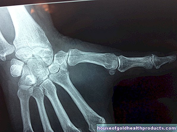
To confirm an ankle fracture, the foot is x-rayed in three planes. A picture is taken from the front, one with 20 degrees of internal rotation and one from the side. Recorded images (pronation / Fricktest) are taken after a fracture has been excluded to check whether the syndesmosis has ruptured.
If the debris is broken, computed tomography (CT) is also required. If a high fibula fracture is suspected, images of the entire fibula are taken in two planes. An additional magnetic resonance tomography (magnetic resonance tomography, MRT) can clarify questionable ligament, soft tissue and cartilage injuries.
Ankle fracture: treatment
The treatment of an ankle fracture depends on the type of fracture: open or closed, postponed or not postponed, Weber or AO classification. The aim of the treatment is to realign the bone fragments and the joint surfaces correctly anatomically and to reconstruct the ligament structures.
If there is a significant displacement and dislocation of the upper ankle joint, the fracture should be closed in an emergency by a doctor at the scene of the accident and immobilized in a suitable splint. This should be done regardless of the subsequent form of treatment, otherwise further soft tissue damage could result.
Ankle fracture: conservative treatment
A stable and non-displaced ankle fracture can be treated conservatively. This is usually a Weber A fracture or Weber B fracture.
The foot is usually immobilized until the swelling has subsided. To do this, the patient first receives a split lower leg cast. After the swelling has subsided, this is replaced by a circular plaster of paris, a plastic splint or a special orthosis (such as Vacoped). Overall, the foot should be immobilized for about six weeks and only partially loaded with about 15 kilograms. Adequate prevention of thrombosis is important, as the foot is not moved for the entire period, which promotes the formation of blood clots (thrombosis).
Even slight irregularities in the upper ankle joint can lead to post-traumatic joint wear (post-traumatic arthrosis). It is therefore important that in the event of an ankle fracture, the foot is aligned exactly anatomically correct - if necessary in an operation. The procedure is best done within the first six to eight hours, if there is no significant swelling. If the soft tissues are swollen, the foot should be stabilized in a fully split and well padded lower leg cast and - until the swelling has subsided - be raised.
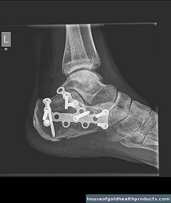
Ankle fracture: surgical treatment
Sprained fractures that cannot be sufficiently immobilized in the cast and tend to shift again, as well as fractures with severe soft tissue damage and multiple injuries, are first stabilized with a joint-bridging "external fixator". This is a special holding system for stabilizing bone fragments. The ankle fracture can thus be immobilized effectively and painlessly. In addition, the fragments can be aligned beyond the ligament structures and the soft tissues can be treated more easily with decongestant measures (such as cold therapy = cryotherapy and pulse compression).
If the capsule and ligaments are affected, they are sutured and pieces of cartilage realigned. The fibula is usually screwed in and stabilized with a neutralization plate. A broken inner ankle is screwed in directly, smaller fragments are fastened with a tension strap.
Fibula fractures up to the middle of the lower leg are directly stabilized. If there is a so-called Maisonneuve fracture, i.e. a high fibula fracture, it is important to realign the ankle joint exactly in terms of length and rotation. To do this, the fibula is immobilized indirectly with a set screw near the ankle joint between the fibula and shin for about six to eight weeks. The ligament structure (syndesmosis) is fixed again with absorbable sutures.
Ankle fracture: follow-up treatment
After the operation of the ankle joint fracture, the foot is held in position with a lower leg split cast. In the case of an "external fixator", the ankle is left in a right-angled position to prevent equinus foot. As soon as the surrounding soft tissues are swollen, the patient is given a special removable shoe (Vacoped) or a circular cast for around four to six weeks - depending on how stable the foot is after the operation and whether ligaments have also been injured.
Ankle fracture: disease course and prognosis
The treatment shows good results in 95 percent of patients with a Weber A fracture. In patients with a Weber C fracture, it is 75 percent.
After an operation, you should not put more than 15 to 20 kilograms on the foot for four to six weeks. Only after six weeks can you put full weight on the ankle again. You can only be active in sports after three to six months. Your doctor will give you more details.
If an implant was used during the operation, it will be removed after about 10 to 12 months. Adjusting screws can be removed after just six weeks.
If a Volkmann fracture is incompletely aligned, early osteoarthritis can develop because the fibula has been incorrectly anatomically aligned or the cartilage damage is too great.
In general, the following applies: The treatment of an ankle fracture shows very good results in 80 percent of all cases if a functional and exercise-stable follow-up treatment is started particularly early.
Tags: drugs menopause teenager