Calcaneal fracture
Dr. med. Mira Seidel is a freelance writer for the medical team.
More about the experts All content is checked by medical journalists.A calcaneus fracture affects the largest bone in the foot - the calcaneus. The cause is usually a fall from a great height or direct force. The typical symptoms are pain in the rear foot and heel area. If the fractions of a calcaneus fracture are not offset against each other, it can be treated conservatively. Otherwise, surgery is usually carried out. Find out more about the calcaneus fracture here.
ICD codes for this disease: ICD codes are internationally recognized codes for medical diagnoses. They can be found, for example, in doctor's letters or on certificates of incapacity for work. S02
Calcaneal fracture: description
The calcaneus fracture is one of the most common tarsal fractures with 60 percent. It affects men between the ages of 25 and 45 most often. A calcaneus fracture often occurs in the context of occupational accidents.
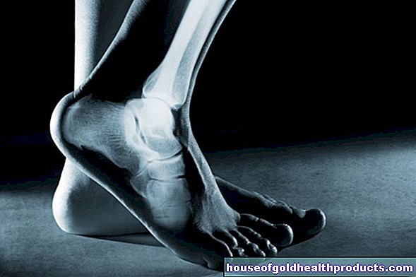
The heel bone (calcaneus) is the largest bone of the foot and is located at the back of the heel. It takes on a central function for the arch of the foot. The shape of the calcaneus is designed for heavy loads. The Achilles tendon attaches to the so-called tuber calcanei at the posterior end of the calcaneus. The bone is connected to the talus via three joint surfaces and to the cuboid bone (cuboid), a tarsal bone, via another joint surface.
Heel fracture: symptoms
The typical symptoms of a calcaneus fracture are pain in the hindfoot and heel area. In addition, there is considerable pressure-sensitive swelling on the lateral rear foot and on the sole of the foot. The rear foot is usually shortened, which makes the contour of the heel look clumsy. Because of the pain, the affected person can no longer put weight on the foot and move the lower ankle joint properly. Furthermore, accompanying injuries to the small muscles of the foot can occur.
In some cases there is an open fracture of the calcaneus. The skin and soft tissues are so damaged that bone fragments may be visible from the outside. An open calcaneal fracture must be treated immediately in an emergency.
Heel fracture: causes and risk factors
The mechanism of an accident in a calcaneus fracture is either a compression from above, as in a fall from a great height, or a direct external force. When falling from a great height, both heel bones are usually broken - this applies to around ten percent of all cases. Multiple injuries often affect the spine or legs.
A calcaneus fracture can also occur if the foot is pinched. This happens, for example, in the context of a car accident when the feet get under the pedals in the car footwell. Another possible cause of a broken heel bone is if the foot becomes twisted.
Sometimes there is also a so-called avulsion fracture of the heel bone: If the calf muscle contracts strongly, the attachment point of the tendon of the fibula muscle on the heel bone (calcaneal tuberosity) can tear out. This break is seen more frequently in people with osteoporosis.
Heel fracture: examinations and diagnosis
A specialist in orthopedics and trauma surgery can best determine whether the heel is broken. The first step is an interview: the doctor asks you exactly how the accident happened and your medical history (anamnesis). Possible questions are:
- What is the exact course of the accident?
- Did you hit your foot or did something fall on your foot?
- Do you have pain?
- Did you already have complaints such as pain and restricted mobility?
This is followed by a physical examination: the doctor examines the foot carefully and pays attention to possible accompanying injuries. He also has to check whether a compartment syndrome has developed: The pressure in a muscle box (= group of muscles that is surrounded by a barely stretchable fascia) has increased due to swelling and blood so that nerves, muscle tissue and vessels are squeezed off. If the tissue is permanently damaged, so-called claw toes can arise.
This is followed by imaging examinations: X-rays of the foot from the side and from above as well as of the upper ankle are taken. A multi-slice computed tomography (CT) scan is usually performed before an operation in order to be able to plan the procedure precisely.
Calcaneal fracture: treatment
Treatment of a broken heel bone is always tailored to the individual patient. The aim of treatment is to restore the adjacent articular surfaces and the structure of the rear foot. Sometimes an operation is necessary for this, but it is technically difficult.
Calcaneal fracture: conservative treatment
Non-displaced breaks are usually treated conservatively. First, the patient is given a plaster splint to keep the foot still. In the lower leg cast or relief shoe (vacuped shoe), which is worn for about 10 to 12 weeks, the injured foot can be partially loaded. After that, slowly increasing, he can be fully loaded again.
Heel fracture: surgical treatment
Surgery is an option if the calcaneus fracture is displaced. The right time for the intervention plays a decisive role: If the heel bone has recently been closed, surgery must not be performed if the surrounding soft tissues are still swollen. The so-called "wrinkle test" is used to determine the correct time for the operation: The operation is only advisable when the skin in the affected region has already swollen enough to wrinkle up. This is usually the case after six to eight days.
A broken heel bone is technically very demanding. If the bone has been dented (impression fracture), this usually requires a so-called spongiosaplasty: This involves inserting the body's own bone material into the bone. The bone parts are held in position by screws or angle-stable plates.
After a fracture of the heel bone - if there is a malalignment or arthritis - an orthopedic mechanic can make individual shoes.
Heel fracture: disease course and prognosis
The healing time and the result of the treatment depend on many factors: age, comorbidities, the physical strain at work and the type of break (postponed or not) all play a role. As a general rule, the healing of a calcaneus fracture is not that easy, because despite optimal treatment, there are often minor functional restrictions. Comminuted fractures in particular lead to considerable difficulties with chronic pain when the foot is put under pressure.
Heel fracture: complications
If a calcaneus fracture is operated on, there is always the risk of impaired wound healing: up to 32 percent of patients struggle with it. The sural nerve, a sensitive nerve in the lower leg, can also be injured during the operation. Another dreaded complication is the compartment syndrome, which occurs ten percent of all cases. Last but not least, premature osteoarthritis of the joint is observed in some patients, caused by the cartilage damage that was associated with the fracture of the calcaneus.
Tags: Menstruation drugs pregnancy birth
