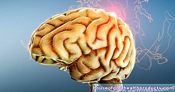
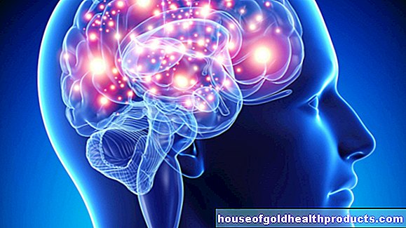
Glioblastoma
Ricarda Schwarz studied medicine in Würzburg, where she also completed her doctorate. After a wide range of tasks in practical medical training (PJ) in Flensburg, Hamburg and New Zealand, she is now working in neuroradiology and radiology at the Tübingen University Hospital.
More about the experts All content is checked by medical journalists.Glioblastoma (glioblastoma multiforme) is a malignant brain tumor. It usually develops within a short period of time in middle-aged people. Risk factors are largely unknown. Despite intensive treatment consisting of surgery, radiation and chemotherapy, the average glioblastoma life expectancy is only a little over a year. Here you can read everything you need to know about glioblastoma.
ICD codes for this disease: ICD codes are internationally recognized codes for medical diagnoses. They can be found, for example, in doctor's letters or on certificates of incapacity for work. D43C71D33

Glioblastoma: description
As a brain tumor, glioblastoma belongs to the group of gliomas. The World Health Organization (WHO) assigns grade 4 glioblastoma to brain tumors. This is the highest degree of severity that a brain tumor can reach.
Most of the time, the tumor forms in one hemisphere of the cerebrum and grows quickly over the bar into the other hemisphere of the brain. Its shape then resembles a butterfly, which is why it is sometimes clearly referred to as a "butterfly glioma".
If you examine the tumor tissue under the microscope, you can see small cavities (cysts), dead tissue (necroses) and hemorrhages. This colorful and often variable appearance has given the tumor the name glioblastoma multiforme or colored glioma.
Primary and secondary glioblastoma
Because its tumor cells are derived from the astrocytes in the brain (a special type of cell), glioblastoma is also known as astrocytoma (grade IV). Depending on the exact origin, a distinction is made between a primary and a secondary type of tumor:
A primary glioblastoma arises directly from healthy astrocytes and is much more common than a secondary tumor. It can develop within a few weeks and primarily affects older people between the ages of six and seven.
A secondary glioblastoma develops from an astrocytoma of lower WHO grade, i.e. an already existing tumor made from astrocytes. In this case, the glioblastoma is the end stage of a prolonged tumor disease. The maximum age of the patients is between 50 and 60 years of age.
Glioblastoma: incidence
Glioblastoma is more common in men than women. Every year in a small city with 100,000 inhabitants such as Trier or Cottbus, around three people develop such a brain tumor. This is not only the most common glioma, but also the most common primary malignant brain tumor in adults.
Special variant: gliosarcoma
Gliosarcoma is a variant of the classic glioblastoma that differs from it in certain tissue properties. Diagnosis, therapy and prognosis are the same for both.
Glioblastoma: symptoms
As with almost all diseases of the brain, the symptoms of glioblastoma also depend primarily on the exact location of the proliferating tissue. Depending on the brain region, completely different symptoms can occur. If a glioblastoma causes symptoms, these usually appear much more suddenly and gain weight more quickly than with other brain tumors. This is because this tumor can develop within a few weeks and grows very quickly. The brain has no way of adapting to other pressure conditions so quickly.
Common glioblastoma symptoms are headaches. They typically appear during the night or early in the morning and get better as the day progresses. Unlike ordinary headaches, they recur regularly and become more and more severe. Medicines often remain ineffective.
Other possible symptoms are seizures and changes in personality. If the glioblastoma grows in the language center or in the control centers of individual muscles, those affected have difficulty speaking or moving. Since these symptoms can occur suddenly, it is not uncommon for a stroke to be misdiagnosed.
In the end-stage glioblastoma, the tumor is usually so large that it causes increased intracranial pressure. Those affected are often nauseous, especially in the morning. Some vomit. If the pressure continues to increase, patients often appear tired or sleepy. In extreme cases, a glioblastoma can even cause comatose conditions.
Glioblastoma Symptoms
You can read more about individual symptoms of brain tumors such as glioblastoma in the article Brain Tumor Symptoms.
Glioblastoma: causes and risk factors
A glioblastoma arises from so-called astrocytes. These cells form the majority of the supporting cells (glial cells) in the central nervous system. They demarcate the nerve tissue from the surface of the brain and the blood vessels. Just like other cells in the body, astrocytes are renewed regularly. Errors can occur that lead to uncontrolled cell growth and ultimately to a tumor.
In addition, a glioblastoma can also arise from an already existing tumor: In an astrocytoma classified as second or third degree by the WHO, the tumor cells can change malignantly, turning it into a grade 4 glioblastoma. This glioblastoma course is less common.Most of the time, the tumor forms directly (primarily), i.e. from healthy cells.
Risk factors for glioblastoma
Why a glioblastoma develops has not yet been adequately clarified. The only established risk factor is ionizing radiation. People are usually only exposed to harmful doses of radiation as part of radiation therapy, i.e. irradiation of another tumor can cause such a brain tumor.
It is also known that people with certain underlying diseases are more likely to develop glioblastoma than those without these diseases. These include, on the one hand, Turcot's syndrome and, on the other hand, diseases that are generally associated with a tendency to form gliomas (such as glioblastoma): neurofibromatosis type I (von Recklinghausen's disease) and type II, tuberous sclerosis (Bournville-Pringle's disease) and Li Fraumeni Syndrome. These very rare diseases are usually associated with typical skin changes.
Glioblastoma: examinations and diagnosis
Symptoms of this brain tumor, such as headaches, speech disorders or epileptic seizures, which usually appear suddenly, cause most patients to see a neurologist. To collect the medical history (anamnesis), he first inquires in detail about the symptoms and their temporal course as well as any underlying or previous illnesses. In order to gain a better overview, the doctor then carries out a neurological examination. If a brain tumor is suspected, he will initiate further investigations.
Magnetic resonance and computed tomography
The most important diagnostic test for glioblastoma is magnetic resonance imaging (MRI) of the skull. If this is not possible for certain reasons (e.g. if you have a cardiac pacemaker), computed tomography (CT) is used as an alternative imaging method. Most of the time, the patient is injected with a contrast agent before the examination, which is then absorbed by the tumor. As a result, it typically appears as a bright, ring-shaped structure in imaging. Although this appearance is very characteristic of a glioblastoma, a tissue sample is usually taken (biopsy).
biopsy
The removal and examination of a tissue sample from the tumor serves, on the one hand, to secure the diagnosis and, on the other hand, to determine precise tissue properties. These can influence the subsequent glioblastoma therapy. If the tumor cells have lost a certain area of the genetic material (1p / 19q) or are chemically changed in a certain gene region (MGMT), they can be better treated with chemotherapeutic agents. Tumors with these changes can therefore be treated in a more targeted manner.
Glioblastoma: treatment
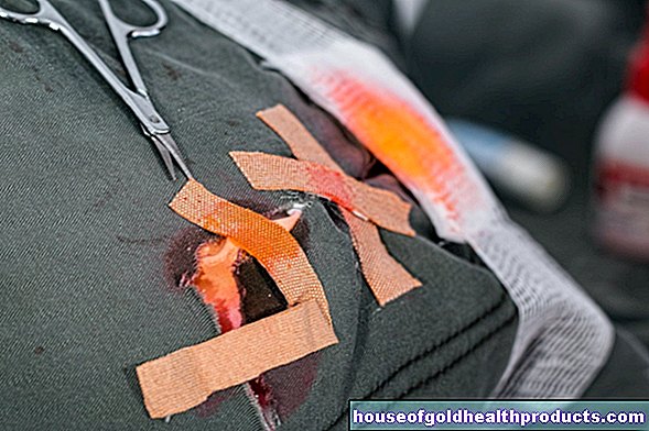
For glioblastoma, the treatment of choice is an operation that is as radical as possible. The tumor region is then irradiated in order to kill any remaining cells of the tumor. At the same time, most patients receive chemotherapy with temozolomide. It will continue for another six months after the irradiation. Elderly patients whose tumors meet certain MGMT properties can also be treated with radiation or chemotherapy alone.
If the tumor returns after successful therapy, a new operation, radiation or chemotherapy will be decided on an individual basis. In addition to temozolomide, active substances such as CCNU or the antibody bevacizumab are available as drugs.
Tumor therapy fields
In addition to surgical, drug and radiation therapy, special treatment centers offer a fourth therapy option with so-called tumor treatment fields (TTFields). These are alternating electrical fields of a certain frequency (200 kHz) that are supposed to inhibit the growth of the glioblastoma. The aim is to maintain the state that has been reached up to that point for as long as possible.
For the TTFields treatment, special ceramic gel pads are stuck to the shaved scalp, creating a kind of hood. Alternating electric fields then build up via these pads. As a result, the tumor cells should no longer be able to divide properly and, at best, die.
The alternating fields are generated by a battery-operated device that the patient can carry in a backpack. In general, this glioblastoma therapy takes place on an outpatient basis - i.e. predominantly in the home or everyday life of the patient. Experts recommend using the TTFields for at least 18 hours a day. The glued-on pads remain on the scalp and are changed approximately every three to four days.
In addition to TTFields treatment, patients continue to take the drug temozolomide.
TTFields will assume the costs
Since May 2020, the statutory health insurances have been taking over treatment with TTFields for patients with newly diagnosed glioblastoma. The prerequisite is that the tumor does not grow again (early) after radiochemotherapy has been completed. To rule out this, doctors first order a magnetic resonance imaging (MRI) scan of the head.
Doctors from various disciplines - such as neurology, radiation therapy and cancer medicine - determine which therapy is particularly suitable for each patient in advance in a so-called tumor conference. Only when the doctors involved recommend the TTFields does the statutory health insurance pay for the treatment.
No standard therapy (yet)
According to the current guidelines of the European Society for Neuro-Oncology, treatment with tumor therapy fields cannot (yet) be regarded as standard therapy for glioblastoma. Some experts, such as the German Society for Neurology (DGN), initially call for further, independent studies. Among other things, these are intended to confirm the promising results of the registration study.
According to this study, patients on combined TTFields and temozolomide therapy lived longer than those who received the drug without concomitant TTFields treatment. The quality of life of the two patient groups was similar in both groups.
Experts from the Institute for Quality and Efficiency in Health Care (IQWiG) rated the additional benefit of the combined therapy as positive in a statement. The Federal Joint Committee (G-BA) followed this assessment and included the treatment option in the service catalog of the statutory health insurance companies.
However, critics consider the results of the registration study for the TTFields add-on treatment to be less clear. For example, they criticize the study design, as there was no comparison group with a “sham treatment”. In the opinion of these critics, the positive contribution of a TTFields treatment is still unclear. Accordingly, the study participants could also have lived longer because they received more intensive care.
possible side effects
Let your doctor explain in detail the advantages and disadvantages of treatment with tumor therapy fields. Overall, the TTFields treatment is considered to be well tolerated. The most common side effects recorded in the registration study were skin irritation from the attached pads (reddening, rarely itching or blisters).
In a statement, the IQWiG experts also point out that some patients may feel restricted in their everyday life through long daily use of the wired pads.
Relief of the symptoms of the disease
In addition to the therapies listed above, which fight the tumor directly, measures to alleviate the symptoms of the disease are often used. Since glioblastoma has a very poor prognosis, the disease is difficult to deal with for many people affected and their families. Some can be supported by psychotherapy or pastoral care.
Glioblastoma: disease course and prognosis
As a rule, no cure for glioblastoma can be achieved even with maximum therapy. Patients who have undergone surgery, radiation, and chemotherapy have a median survival of approximately 15 months. Almost ten percent of patients survive five years. Without therapy, the mean survival time is around two months. With surgery alone it is around five months, with surgery plus radiation therapy around 12 months.
Life expectancy and quality are also subject to individual factors. The tumor cells do not have the same properties in every person affected. Some are easier to treat than others. If the tumor shrinks quickly during therapy, the glioblastoma prognosis is usually better than in other cases.
In addition, chemotherapy drugs and radiation therapy are tolerated differently by patients. If the side effects are too strong, glioblastoma therapy does more harm than good to those affected. Then it should be individually weighed whether the treatment should be continued less intensively. In this way, those affected influence the glioblastoma course themselves to a certain extent: They accept a shorter lifetime if this improves their quality of life with the glioblastoma.
Tags: drugs sleep drugs