Emphysema
and Sabine Schrör, medical journalistDr. med. Julia Schwarz is a freelance writer in the medical department.
More about the expertsSabine Schrör is a freelance writer for the medical team. She studied business administration and public relations in Cologne. As a freelance editor, she has been at home in a wide variety of industries for more than 15 years. Health is one of her favorite subjects.
More about the experts All content is checked by medical journalists.In the case of emphysema, the alveoli are partially overstretched and destroyed. The result is that the body can no longer be adequately supplied with oxygen. Typical symptoms of emphysema are therefore shortness of breath, shortness of breath and reduced performance. Smoking is the leading cause of lung hyperinflation. What other causes are there, what the consequences of pulmonary emphysema are and what the treatment and prognosis look like, read here!
ICD codes for this disease: ICD codes are internationally recognized codes for medical diagnoses. They can be found, for example, in doctor's letters or on certificates of incapacity for work. J44J43

Brief overview
- What is emphysema? Chronic lung disease associated with overinflation and destruction of alveoli. As a result, the body is increasingly poorly supplied with oxygen.
- Causes: Smoking, chronic inflammatory processes (chronic bronchitis, COPD, etc.), alpha-1 antitrypsin deficiency, old age, inhalation of pollutants (dust, gases, etc.), scarring in the lung tissue
- Symptoms: shortness of breath (at first only during physical exertion, later also at rest), cough, reduced performance, increased susceptibility to infections. In the advanced stage also blue lips and nails (cyanosis), barrel-shaped chest (barrel chest), poor general condition with muscle breakdown, right heart failure (type of heart failure).
- Examinations: initial consultation (anamnesis), physical examination, x-ray, computed tomography, blood gas analysis, lung function test
- Treatment: cessation of smoking, breathing training, inhalation of saline solution, adequate fluid intake, drug therapy, with advanced pulmonary emphysema possibly long-term oxygen therapy, surgery. Lung transplantation in very severe cases. Vaccinations against pneumococci and flu viruses make sense due to higher susceptibility to infections, treatment of acute respiratory infections with antibiotics.
- Prognosis: No cure possible. Consistent treatment (especially stopping smoking) can slow down or stop the progression of the disease. Possible complications are pneumothorax (collapsed lung), right heart failure with water retention in the legs (edema), jammed neck veins, liver cirrhosis.
Pulmonary emphysema: knowledge & causes
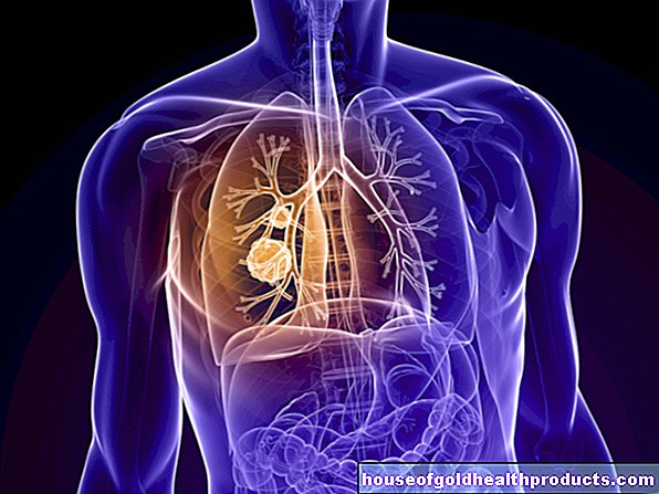
With emphysema, more and more alveoli are pathologically inflated and destroyed. Doctors therefore also speak of overinflation of the lungs.
The alveoli are the smallest structural units of the lungs and are surrounded by a dense network of very fine blood vessels (capillaries). This is where the gas exchange takes place: With every breath, air flows through the windpipe into the alveoli. There the oxygen from the air reaches the blood through the walls of the alveoli. At the same time, carbon monoxide (CO2) is released from the blood into the air in the alveoli. This stale air is then exhaled.
In the case of emphysema, the gas exchange no longer works properly: In those affected, the walls of the alveoli lose their elasticity. The air that flows in with each breath causes the alveoli to be overstretched. After all, they can even burst. Adjacent destroyed alveoli then combine to form larger bubbles that collapse when you exhale. By overinflating the alveoli, the smallest bronchi (bronchioles) are narrowed and also collapse easily. All of this makes it difficult to breathe out: Patients can no longer fully breathe out the air they breathe. Part of it remains in the vesicles that are still present, so that when you breathe in there is less space for new, oxygen-rich air. The result is an increasing lack of oxygen.
This is what happens with emphysema
In the case of emphysema, the wall structure of the alveoli is irrevocably destroyed. This leads to a sack-shaped expansion of the air spaces.
Pulmonary emphysema: causes
Certain proteins usually play a decisive role in the development mechanism of pulmonary emphysema:
Every time you inhale, pathogens and other harmful substances enter the lungs with the air. The immune system is normally prepared for this: Defense cells in the alveoli regularly release protein-degrading proteins, so-called proteases. They can render invasive germs and foreign substances harmless. However, proteases do not differentiate between foreign and own tissue. They can therefore attack and break down the sensitive lung tissue (more precisely: the elastic fibers in the walls of the alveoli). To prevent this from happening, an appropriate amount of protective proteins, known as protease inhibitors, is usually released. The most important representative of these is alpha-1-antitrypsin.
The balance between proteases and protease inhibitors can be disturbed by smoking, chronic inflammatory processes and an alpha-1 antitrypsin deficiency so that lung tissue is increasingly broken down and pulmonary emphysema develops. Other possible causes include repeated inhalation of pollutants, old age, and scar tissue in the lungs.
smoking
Smoking is the leading cause of emphysema. It works by blocking the protective protease inhibitors (such as alpha-1 antitrypsin). These cannot prevent the proteases from attacking the alveoli - pulmonary emphysema develops.
Chronic inflammatory processes
Chronic inflammatory processes in the lungs, like smoking, can cause the destructive proteases in the alveoli to gain the upper hand. In this way, both chronic bronchitis and chronic obstructive pulmonary disease (COPD) can pave the way for emphysema.
Alpha-1 antitrypsin deficiency
In about one percent of patients, pulmonary emphysema develops on the basis of a genetic deficiency in alpha-1-antitrypsin, the main representative of protease inhibitors. As a result, those affected are more susceptible to emphysema than the general population. How high the risk of the disease is in each individual case depends on the severity of the hereditary disease. Pulmonary emphysema develops particularly easily if people with a congenital alpha-1 antitrypsin deficiency also smoke or have a chronic lung disease (such as COPD).
A congenital alpha-1 antitrypsin deficiency can have other consequences in addition to pulmonary emphysema. This includes an increasing destruction of liver cells (liver cirrhosis).
age
The elasticity of connective tissue generally decreases with age. This also reduces the elasticity of the septum walls of the alveoli. This rarely leads to the development of age-related emphysema. This so-called emphysema of old age is not a disease, but a consequence of the natural aging process. Those affected usually show no symptoms. Therefore, the emphysema of old age usually does not have to be treated.
Scar tissue
After surviving pneumonia or tuberculosis, as well as lung surgery (such as the removal of a lung), scar tissue often forms in the lungs. This is more unstable than normal lung tissue and is therefore more easily overstretched. This creates what is known as scar emphysema.
Substances irritating to the respiratory tract
Pulmonary emphysema is rarely caused by inhalation of irritating gases or dust. These inhalative noxae include:
- quartz-containing dust
- Cotton and grain dust
- Welding smoke
- Gases such as ozone or chlorine gas
General air pollution in large cities can also promote emphysema. This is especially true for people who already have another lung disease.
Types of emphysema
There are different types of emphysema. The cause of the lung overextension plays an important role in the classification:
- Centrilobular emphysema: About a third of chronic emphysema patients suffer from a centrilobular form. The upper lung fields are mainly affected, while the lower ones are intact. Doctors attribute this to the fact that pollutants such as cigarette smoke first reach and damage the upper lung tissue.
- Panlobular pulmonary emphysema: This type of pulmonary emphysema is mostly genetic, i.e. it is based on the congenital alpha-1-antitrypsin deficiency described above. The alveoli of the lower lung are particularly affected here.
- Overstretch pulmonary emphysema: If part of the lungs has to be surgically removed, the remaining lung in some cases expands excessively - it creates overstretch emphysema. Compared to the other two types of emphysema, it usually causes fewer symptoms, since the walls of the alveoli have not previously been damaged by chronic inflammation (for example caused by cigarette smoke).
Pulmonary emphysema: symptoms
Symptoms of emphysema develop slowly and insidiously. The type and severity of symptoms depend on the stage of the disease.
First symptoms of emphysema
In the early stages, emphysema patients usually suffer from shortness of breath during physical exertion. In addition, there are sporadic coughs and reduced performance. Many of those affected get tired quickly even with light exertion.
Frequent infections of the respiratory tract such as bronchitis and pneumonia are typical accompanying symptoms of emphysema. This susceptibility to infection results from the increasing destruction of the ciliated epithelium, which lines a large part of the airways. The ciliated cells resemble tiny hairs. They move in waves and thus transport tiny foreign matter particles out of the lungs (mucociliary clearance). Tobacco smoke and other pollutants destroy the ciliated epithelium in the long term, which disrupts the cleaning mechanism. That makes infections easier.
Advanced emphysema
People with advanced emphysema show shortness of breath even at rest, i.e. even without physical exertion. In addition, many patients suffer from a cough with mucous sputum, especially if they have chronic bronchitis at the same time.
The shape of the rib cage can change over time due to the increasing overinflation of the lungs: In the case of pulmonary emphysema, the chest muscles are more stressed when breathing and remain in the inhalation position for a long time. The ribs then run horizontally instead of sloping downwards, and the chest appears "barrel-shaped" (barrel chest). The two pits in the skin above the collarbones disappear - they also appear over-inflated for many of those affected.
Often the general condition of the patient also deteriorates. As a result of the shortness of breath, those affected move less, so that the body breaks down muscle mass. This can make the shortness of breath worse.
The persistent lack of oxygen in the blood is often shown by blue lips and fingers (cyanosis). In addition, the inflated lungs can put excessive strain on the right half of the heart. A particular form of heart failure (heart failure) develops: right heart failure. The overloaded right half of the heart cannot adequately transport the inflowing blood. It backs up in the body's circulation. This blood congestion shows up in bulging neck veins, among other things. Other signs of right heart failure are water retention in the legs (edema).
Two extremes: pink puffer and blue bloater
Pulmonary emphysema patients are divided into two types: the "pink buffer" and the "blue bloater". Both are extreme forms that are rarely observed in full. Most of the time, the transitions are fluid.
Typical features of the Blue Bloater are:
- Obesity
- Pronounced cyanosis (blue discoloration of the lips and fingers, hence the term "blue bloater")
- severe cough with sputum
- Barrel chest
- slight shortness of breath
- "Lip brake" (exhale through loosely placed lips)
Typical features for the pink puffer are:
- Underweight
- no cyanosis due to heavy breathing, rather pale skin color
- pronounced shortness of breath
- dry cough without expectoration
To put it simply: the “Pink Puffer” fights against its pronounced shortness of breath by breathing consciously and intensely. As a result, his blood is sufficiently saturated with oxygen so that cyanosis does not develop. The “blue bloater”, on the other hand, gets used to the chronic oxygen deficiency over time, which often leads to right heart failure due to a so-called cor pulmonale. The right hemisphere of the heart is exhausted because it has to pump against too great a resistance in the pulmonary circulation.
Pulmonary emphysema: treatment
There is no cure for emphysema. This means that the pathological changes in the lung tissue are irreversible. With the right therapy, however, the course of the disease can be slowed down or even stopped.
Do not smoke!
Patients with emphysema should stop smoking immediately and permanently. Some manage to quit smoking all by themselves. But many of them need help. After all, nicotine addiction is one of the strongest addictions. So do not hesitate to seek help in quitting smoking. This can be behavioral therapy or a self-help group. Nicotine substitutes such as nicotine patches, chewing gums or sprays as well as acupuncture or hypnosis can also help with weaning.
Vaccinate
Pulmonary emphysema patients are more prone to respiratory infections. That is why doctors recommend the pneumococcal vaccination and the flu vaccination. The vaccination protection of the pneumococcal vaccination lasts for about five years and should then be renewed. You have to get vaccinated against influenza every year, as the flu viruses are constantly changing.
Treat infections early
Fever and cough with yellow-greenish sputum suggest a bacterial respiratory infection. If pulmonary emphysema patients show symptoms like this, they should definitely seek antibiotic treatment from a doctor. This can prevent a serious course of the disease. Long-term antibiotic treatment is rarely necessary, which the family doctor then has to monitor closely.
Medical therapy
The drugs used for emphysema depend on the stage of the disease and the severity of the symptoms. In principle, the following active ingredients are available (in principle the same as in the treatment of asthma and COPD):
- Beta-2 sympathomimetics: have a bronchodilator effect and are inhaled; there are short-acting (such as salbutamol, reproterol) and long-acting representatives (such as salmeterol)
- Anticholinergics: they too have a bronchodilator effect and are inhaled; Example: ipratropium
- possibly glucocorticoids ("cortisone"): anti-inflammatory effect; are also mostly inhaled; cortisone tablets may only be given in severe cases
In the case of a congenital alpha-1 antitrypsin deficiency, the missing protein can also be replaced with medication. Such alpha-1-antitrypsin substitutes are regularly administered as an infusion.
Long-term oxygen therapy
Patients with severe emphysema often receive long-term treatment with oxygen: pure oxygen is inhaled through a mask for at least 16 hours a day. This can improve the prognosis and improve the quality of life of those affected.
However, not all patients benefit from oxygen therapy. If the body has already got used to an increased C02 level in the blood, the strongest respiratory drive no longer applies: the rising C02 content. Then the lack of oxygen remains as the only respiratory drive. If oxygen is now supplied in an uncontrolled manner, this last respiratory drive is also omitted. Patients stop breathing on their own and may develop carbon monoxide anesthesia. That is why the doctor decides very carefully whether oxygen therapy for pulmonary emphysema makes sense in individual cases or not.
Under no circumstances should you smoke during oxygen therapy, as oxygen is highly explosive. A little glow or a small spark is enough to ignite the gas!
Physical emphysema therapy
With breathing therapy, patients learn special techniques that make it easier to breathe out.Strong abdominal muscles are also helpful for correct breathing technique. Therefore, the therapy also includes targeted abdominal muscle training. In addition, those affected should drink enough and regularly inhale with a saline solution to help cough up the sputum.
surgery
If you have severe emphysema, surgery can be useful. The doctor removes the functionless, over-inflated lung tissue (volume reduction therapy). As a result, the healthy lung tissue is better ventilated again.
In patients with very advanced pulmonary emphysema, the last treatment option is often a lung transplant.
Pulmonary emphysema: examinations and diagnosis
The right person to contact if you suspect pulmonary emphysema is a pulmonologist (pulmonologist). He will first talk to you in detail to collect your medical history (anamnesis). Possible questions during this interview are:
- Do you suffer from shortness of breath?
- Do you cough often? Is the cough dry or associated with sputum?
- Do you have any other complaints?
- Do you smoke? If so, how much and how long?
- How many stairs can you climb without taking a break?
- Do you already have a lung disease (asthma, chronic bronchitis, etc.)?
- Do you have relatives with emphysema, COPD or alpha-1 antitrypsin deficiency?
Physical examination
The anamnesis is followed by a physical examination. The doctor can recognize typical changes that can indicate pulmonary emphysema. This includes, for example, the so-called barrel chest: a barrel-shaped rib cage suggests permanent lung overinflation. This is a clear distinguishing feature of emphysema.
Another characteristic feature is the instinctively performed lip brake on exhalation. Those affected breathe out through loosely placed lips. This breathing technique makes it easier to breathe out because it increases the air pressure in the bronchi.
Bluish discolored lips and fingers are also serious signs of emphysema. The same applies to the so-called drumstick fingers and watch glass nails. The fingers are blown up on the end links like drumsticks, while the fingernails are strongly rounded.
Swollen legs and bulging neck veins are also important signs. They indicate that the right half of the heart is being stressed more. The reason for this can be emphysema.
When listening to the lungs with a stethoscope (auscultation), dry rattling noises (humming or whistling) can often be heard in the case of emphysema. When tapping the chest (percussion) it sounds loud and hollow. The reason is the increased amount of air in the inflated lungs. In contrast, the heart sounds can often only be heard very softly with the stethoscope due to the increased volume of the lungs.
Further investigations
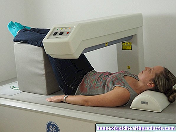
To assess how far the emphysema has progressed, the doctor can X-ray the chest (chest X-ray): The excess air "trapped" in the alveoli can press the diaphragm down (flatten) and appear as a dark area on the X-ray appear. In addition, the vessels, which are normally clearly visible, are often difficult to see (vascular rarefaction).
With a computed tomography (CT) the pulmonary emphysema can be shown in more detail.
With the help of a blood gas analysis and a lung function test, the doctor can determine whether a pulmonary emphysema patient is more likely to be pink buffer or blue bloater. These diagnostic methods show whether only the oxygen level in the blood is reduced (typical for the Pink Buffer) or whether the CO2 level is also increased (typical for the Blue Roater).
Pulmonary emphysema: disease course and prognosis
Pulmonary emphysema is not curable. However, if you stick to the treatment plan prescribed by your doctor, you can slow down or even stop the course of the disease.
One of the most important therapy building blocks is the immediate, absolute and permanent cessation of smoking!
Pulmonary emphysema: complications
Consistent therapy is also important in view of the complications that progressive emphysema can bring: pneumothorax and right heart failure.
Pneumothorax
A serious acute complication of emphysema is the pneumothorax: Normally there is negative pressure in the gap between the lungs and pleura (pleural gap). If, however, an emphysema-damaged alveolus bursts, air can penetrate the gap and remove the negative pressure - with serious consequences: the lungs collapse on the affected side. The collapsed part is no longer ventilated and can therefore no longer take part in the gas exchange. A spontaneous pneumothorax usually manifests itself as a sudden, sharp pain and shortness of breath.
Right heart failure
Pulmonary emphysema increases the pressure in the pulmonary vessels over time. The right half of the heart has to pump against the increased resistance in the pulmonary vessels, which puts increasing strain on it. Over time, this can lead to a right heart failure (right heart failure). The weakened right half of the heart (cor pulmonale) can no longer pump sufficiently, so that fluid collects in the legs (edema). In addition, cirrhosis of the liver and jammed neck veins can occur.
To avoid these complications, you should strictly adhere to the therapy guidelines of your treating doctor and, above all, stop smoking. Then there is a good chance of stopping the emphysema.
Additional information
Self-help groups:
- Patient organization pulmonary emphysema-COPD Germany: https://www.lungenemphysem-copd.de
- COPD-Germany e.V .: https://www.copd-deutschland.de/