Scoliosis
Florian Tiefenböck studied human medicine at the LMU Munich. In March 2014, he joined as a student and has supported the editorial team with medical articles ever since. After receiving his medical license and practical work in internal medicine at the University Hospital Augsburg, he has been a permanent member of the team since December 2019 and, among other things, ensures the medical quality of the tools.
More posts by Florian Tiefenböck All content is checked by medical journalists.In scoliosis, the spine curves to one side. Most of the time, the vertebral bodies are also twisted. Symptoms usually only arise when there is a severe curvature of the spine. Mild forms can often be treated with physiotherapy and a special brace, while severe cases require surgery. Read everything about the causes, diagnosis and therapy of scoliosis here.

Brief overview
- Definition: permanent lateral curvature of the spine
- Frequent symptoms: shoulders of different heights, crooked pelvis, crooked head, lateral "rib hump", back pain, tension
- Possible consequences: stiffening of the respective vertebral section, early wear
- Examinations: Physical examination, Adams test, mobility / strength tests, X-rays, determination of skeletal maturity
- Treatment: physiotherapy, corset, plaster cast, clamp technique, surgery
Scoliosis: description
In order to understand what constitutes scoliosis, one must first know how a healthy spine is structured.
Structure of the spine
A healthy spine consists of about 33 vertebrae: seven cervical vertebrae, twelve thoracic vertebrae, five lumbar vertebrae, five fused sacral vertebrae and about four - also fused - coccyx vertebrae. The vertebral bodies are connected to the neighboring vertebrae and ribs via bone processes.
When viewed from the side, the spine is shaped like a double "S". The cervical and lumbar spine bulge forward (lordosis), the thoracic and sacral spine (sacrum) backward (kyphosis). If you look at the spine from behind, it forms an almost straight line with its spinous processes from the head to the anal fold. The vertebral bodies lie evenly on top of one another and between each two of them there is an intervertebral disc as a shock absorber.
The spine is an important part of the supporting skeleton and also protects the spinal cord, a bundle of nerve tracts that carry signals between the body and the brain.
Spinal segments
What is Scoliosis?
In scoliosis, the structure of the spine is disturbed. The name of the disease is derived from the Greek word skolios, which means "crooked": In this case, the spine not only curves forwards and backwards, but also to the side.
According to the guideline-compliant definition of scoliosis, this clinical picture is "a permanent (fixed) lateral curvature of the spine by at least ten degrees Cobb's angle. This angle indicates how strong the lateral curvature of the spine is and can be based on an X-ray image Depending on which side the spine curves on, doctors speak of right or left convex scoliosis.
Measurement of scoliosis with the COBB angle

In addition, the individual vertebral bones and the entire spinal column are twisted in their longitudinal axis (rotation and torsion). This shows the bony vertebral body processes (spinous process, spinous process). The side of the appendages that points towards the abdomen or the chest rotates in the direction of the curvature of the spine. The rotation is strongest at the apex of the scoliosis and decreases again at the ends of the crooked spine.
As scoliosis progresses, the corresponding section of the vertebra can stiffen.
Due to the varying degrees of twisting, tension and pressure forces arise between the individual vertebral bodies. As a result, the vertebral bone also has a distorted bone structure (torqued): the vertebral body is higher on the outwardly curved side than on the inwardly facing side. The same applies to the intervertebral discs between the vertebral bones. This results in permanent crooked growth. Experts also refer to the twisted and crooked spine as torsional scoliosis.
Torsional scoliosis usually only occurs on the main curvature. In order to compensate for severe scoliosis, muscle strength creates secondary curves in the spine in the direct vicinity of the main curve (static compensation). However, the minor curvatures have no rotation or torsion. If it does, it's called multiple scoliosis.
What types of scoliosis are there?
Scoliosis can be divided into different forms, depending on the point of view. For example, a general distinction is made between idiopathic scoliosis and secondary scoliosis.
- idiopathic means that no specific trigger for the disease can be found.
- Secondary scoliosis, on the other hand, is always the result of a known cause.
These “real” (structural) scolioses are to be distinguished from a scoliotic malposition (also functional scoliosis).
Scoliotic bad posture is temporary and normalizes again through passive or active movements. It arises, for example, to compensate for a pelvic inclination.
Since in many cases the cause of scoliosis is not known, it cannot be effectively prevented.
Real scolioses can be further differentiated according to age and curvature pattern:
Scoliosis of different age groups
Among other things, scolioses can also be differentiated according to the time of first occurrence. The early form is called infant scoliosis, and in most cases it resolves without therapy. Doctors speak of infantile scoliosis if the curvature of the spine occurs by the age of three. Scoliosis in children between the ages of four and ten is known as the juvenile form.
However, adolescent scoliosis is most common from the age of eleven. The spine is usually curved to the right in the thoracic area (right convex scoliosis). Girls are more often affected than boys.
Scoliosis curvature pattern
In addition, scoliosis can be assigned to the middle (or vertex) of its main curvature in the spine. In thoracic scoliosis, the curvature is in the thoracic spine (thoracic spine). Thoraco-lumbar scolioses have their greatest lateral curvature where the thoracic spine merges into the lumbar spine (LWS). A curvature of the spine in the lumbar area is called lumbar scoliosis.
- In some cases, those affected suffer from thoracic and lumbar spine scoliosis. A curvature pattern is formed which - if you look at the patient's back from behind - is reminiscent of the letter "S" (double arched).
- If the spine is completely bent to one side, doctors speak of a C-shaped scoliosis.
- If the spine curves in all sections (thoracic spine, lumbar spine and their transition) alternately to the right and left, a double S spine, also known as triple scoliosis, is created.

Scoliosis degree of curvature
Scoliosis can also be classified according to how severely curved the spine is:
- mild scoliosis: angle up to 40 degrees (1st degree scoliosis)
- Moderate scoliosis: angle between 40 and 60 degrees (2nd degree scoliosis)
- severe scoliosis: angle of 61 to 80 degrees (3rd degree scoliosis)
- very severe scoliosis: angle over 80 degrees (4th degree scoliosis)
Scoliosis Frequency: How often does the disease occur
Around two to five percent of the population suffer from idiopathic scoliosis. According to a study by the Maimonides Medical Center (USA), the incidence in old age (60 to 90 years) can increase up to 68 percent.
The greater the curvature of the spine and the higher the age, the more often women and girls are affected. Mild scoliosis can be found in boys. More pronounced scolioses, with a Cobb angle greater than twenty degrees, are about seven times more common in women than in men.
-
Scoliosis therapy - the earlier, the better!
Three questions for
Dr. med. Frank Meinhard Balensiefen,
Specialist in Orthopaedics -
1
How do I recognize scoliosis?
Dr. med. Frank Meinhard BalensiefenThere are various clues. Especially if a family has a history of scoliosis, you should be vigilant. In children and adolescents, for example, the gait pattern changes. Differences in leg length or a noticeably rounded back, known as a seat hump, can also indicate scoliosis. Sports teachers and trainers often have a good eye for abnormalities in the spine.
-
2
Why is early treatment so important in children and adolescents?
Dr. med. Frank Meinhard BalensiefenThe sooner the treatment starts, the better. This means that the young patient's growth phase can be optimally used and measures such as a corset or targeted training of the back muscles can be carried out over a long period of time. After the pubescent growth spurt, it is much more difficult to achieve satisfactory results with the help of conservative therapy.
-
3
When does an operation make sense?
Dr. med. Frank Meinhard BalensiefenConservative measures like physical therapy, exercise, and breathing muscle training can keep scoliosis from worsening. Sometimes this can avoid surgery. However, if the vertebral body is very bent to the side by more than 30 degrees, surgery is advisable. Especially when the function of internal organs such as the heart and lungs are restricted as a result.
-
Dr. med. Frank Meinhard Balensiefen,
Specialist in OrthopaedicsHead of the Endo Center at the Munich East Orthopedic Center, hip and knee arthroplasty, pediatric orthopedic diseases, sports medicine, hand and foot surgery, since 1997 doctor in charge of the German national ice hockey teams

Scoliosis: symptoms
In a large number of cases, scoliosis is a purely cosmetic problem. However, the longer it is left untreated, the more likely it is that pain will develop as the disease progresses. How pronounced the symptoms are always depends on how advanced the curvature is.
Cosmetic scoliosis symptoms that can be seen with the naked eye include, among others
- shoulders at different heights
- oblique pelvis or pelvis protruding on one side
- head held crooked
With pronounced scoliosis, the so-called rib hump often occurs, muscle bulges can form in the lumbar and neck area.
Due to increasing signs of wear and tear, those affected have more problems with muscle tension and pain, especially from the middle of the third decade of life. Lung capacity can also decrease and you may experience shortness of breath, a feeling of pressure in the chest or a racing heart.
Read everything you need to know about scoliosis symptoms here.
Scoliosis: causes and risk factors
About 90 percent of all scolioses are idiopathic, so you don't know why they arise. For the remaining ten percent - secondary scoliosis - there are various possible causes that lead to a curvature of the spine.
Malformation scoliosis
This form of scoliosis is due to congenital malformations of individual parts of the spine, for example
- wedge-shaped vertebral bodies (different rim heights)
- split or semi-formed vertebral bones
- congenital malformations of the ribs (synostoses)
- Defects in the spinal canal (e.g. diastomatomyelia)
Experts therefore refer to it as congenital scoliosis.
Myopathic scolioses
These spinal curvatures are caused by muscle diseases (including hereditary muscle weakness diseases). The most common is Duchenne muscular dystrophy, in which a certain muscle protein is not formed. As a result, children suffer from increasing muscle weakness and wasting at an early age. More than half of all those affected develop scoliosis in the course of Duchenne muscular dystrophy, mostly in early adolescence and after they have lost their ability to walk.
Arthrogryposis can also lead to severe scoliosis in severe cases. It is a congenital joint stiffness caused by changes in the tendons, muscles and connective tissue.
Neuropathic Scolioses
In this form, damage to the nervous system leads to a crooked spine. Muscles that stabilize the spine (abdominal and back muscles) then no longer function as usual. This creates an imbalance, the spine curves in the direction of the flaccid muscles.
Among other things, these diseases of the nervous system lead to scoliosis:
- Myasthenia gravis (muscle paralysis)
- viral inflammation of the spinal cord (myelitis)
- early childhood brain damage (e.g. infantile cerebral palsy)
- neurodegenerative diseases with damage and loss of nerve cells (e.g. spinal muscular atrophy with a decrease in the second nerve pathway to the muscles)
- Cave formation in the spinal cord due to cerebral water congestion (syringomyelia)
- malignant or benign growths (e.g. spinal tumors)
Other causes of scoliosis
In addition to the muscle and nerve diseases mentioned, numerous other clinical pictures can be associated with scoliosis of varying degrees. The surrounding connective tissue, but mostly also the bone and cartilage structures, are directly affected. The table gives some examples.
|
Disease group |
Causes of Scoliosis (Examples) |
|
Connective tissue disorders |
|
|
Rheumatic diseases |
|
|
Malformations of the bone-cartilage structures (osteochondrodysplasia) |
|
|
Bone infections (acute, chronic) |
|
|
Metabolic diseases (metabolic disorders) |
|
|
Lumbar sacral changes in the lumbar vertebrae and sacrum area |
|
Accidents can also lead to scoliosis. These post-traumatic scolioses occur, for example, after a vertebral bone fracture, burns or spinal cord injuries. Furthermore, some medical interventions cause a curvature of the spine, such as radiation or laminectomies. In the latter, part of the vertebral bone (vertebral arch, possibly with a spinous process) is removed.
As with many diseases, experts suspect that scoliosis can also be inherited. In 97 percent, there was a higher incidence of family members. Among identical twins, up to 70 percent of the time, both of them develop scoliosis. Since scoliosis increases with age, researchers assume that ultimately signs of wear and tear also have a decisive influence (degenerative changes).
Scoliosis: diagnosis and examination
The specialist in diseases of the musculoskeletal system is the orthopedist. There are also pediatricians and pediatric orthopedists who specialize in scoliosis. First, the doctor collects the medical history (anamnesis) and asks the patient or their caring relatives, among other things, the following questions:
- When did you first notice the crooked spine?
- Do you suffer from complaints such as back pain?
- Have you already had your first menstrual period (menarche) or your voice breaking?
- How fast have you grown over the past few years?
- Do you have any other known illnesses, such as deformity of the feet, a crooked pelvis, muscle or nerve diseases?
- Do you have any known cases of scoliosis in your family?
The US American Scoliosis Research Society regularly publishes questionnaires for patients suffering from scoliosis (current version SRS-30). The German translation of the questionnaire is also used by doctors in this country.
Tip: Those affected should fill out the questionnaire at regular intervals. This allows them to indicate how they perceive the course of the disease and assess the success of the therapies carried out.
Physical examination
After the interview, your doctor will perform a physical examination. First he determines the standing and sitting size, then he assesses the back and especially the shape of the spine. If the line of the spinous processes deviates, he will notice a so-called overhang. The chest is shifted to the side. In the case of scoliosis, a straight line from the last cervical vertebra down no longer ends in the anal fold but next to it.
He also checks the equality of the shoulder blades (symmetrical shoulder stand) and the waist as well as the outline of the torso. With scoliosis, the shoulders are of different heights. The two so-called waist triangles are also of different sizes, i.e. the distances from the left and right hanging arm to the torso.
During the physical examination, the doctor also looks at the still image from the side. This enables him to recognize excessive hunching (hyperkyphosis) or a spine that is strongly curved towards the abdomen (hyperlordosis, e.g. hollow back).
In rare, pronounced cases, a distinct thoracic vertebral hump is formed. The thoracic spine is then not only curved to the side, but also strongly backwards (kyphoscoliosis).
Such kyphoscoliosis usually occurs with other diseases, for example rickets, bone marrow inflammation or tuberculosis of the vertebral bodies.
In addition, a crooked pelvis or legs of different lengths can be noticed in the context of scoliosis (leg length difference).
The doctor will also examine the skin of the back, as diseases of the spinal cord can already show up here. Light brown and even spots on the skin, so-called café-au-lait spots, on the other hand, are typical of the hereditary disease neurofibromatosis type 1 (Recklinghausen's disease), which mainly affects the skin and the nervous system. Here, too, those affected can suffer from scoliosis, especially kyphoscoliosis.
Physical examination in the infant
Scoliosis in infants is revealed by various posture tests. For example, if the child lies with their stomach on the examiner's hand, they can easily see a crooked spine, as the curvature is usually clearly visible on the back. Differences in arm and leg development can be seen in the Vojta side tilt reaction. To do this, the doctor holds the child sideways and pays attention to the infant's body tension. On the side facing away from the curvature, the body usually falls significantly more slack than on the side towards which the curvature is directed.
Scoliosis is also clearly visible in the vertical hanging reaction according to Peiper and Isbert. Hanging upside down by the feet, the entire baby's body is curved in a C-shape to one side.
Adams test
During this examination, the doctor asks you to bend forward with your knees extended. If he now looks at your back, he may be able to recognize typical signs of scoliosis, for example a rib hump on the back with a bent posture or muscle bulges in the neck and lumbar area.
As a rule, the doctor measures the shape of the rib hump or the muscle bulges using a so-called scoliometer or inclinometer. He compares the heights of the left and right side with each other. According to the guidelines, deviations of more than five degrees are to be regarded as pathological. In these cases, further examinations follow, in particular x-rays of the spine.
Examination of mobility, strength, flexibility and reflexes
As part of the physical exam, the doctor will also ask you to lean back and forth and to the side. In this way, he checks the mobility of the spine. He also measures the finger-floor distance in a position bent forward to the maximum with straight legs. Ideally, you can touch the floor (0 cm), but this is rarely possible with pronounced scoliosis. In addition, the doctor will check whether the curvature of the spine can be actively compensated for by your own movements or by manual assistance from the doctor (passive, manual redressability). “Real” structural scoliosis can hardly be changed, if at all.
It is also important to recognize any abnormalities in the nervous system that may cause scoliosis or a curvature of the spine or hereditary connective tissue diseases (Marfan syndrome).
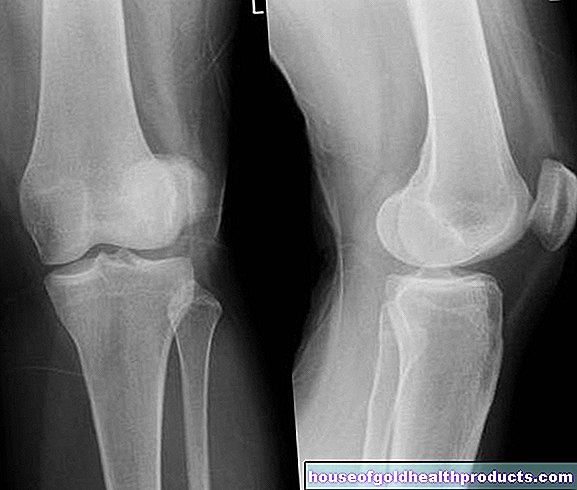
roentgen
In many cases, the doctor can diagnose scoliosis based on the physical exam alone. However, if a spinal curvature is suspected, he will always arrange for an X-ray examination. The whole spine is depicted while standing, viewed from the front (or back) and from the side.
With the help of the X-ray images, the doctor can measure the Cobb angle (in infant coliosis it is more likely the angle of departure of the ribs RVAD), determine the major and minor curvatures, identify the vertebrae and the terminal vertebrae and determine the curvature pattern. This procedure is important for the subsequent scoliosis therapy. In addition, malformations or deformations of the bones can be detected in this way.
X-ray scoliosis
Determination of skeletal maturity
In order to be able to assess the course of scoliosis in adolescents, it is important to determine the level of spinal growth. For this purpose, the skeletal maturity is assessed using x-ray images based on the ossification of the iliac crest processes (apophyses). These processes ossify more and more with increasing age; if they are completely ossified and the apophyses are closed, skeletal growth is complete. The bone age can also be determined using an X-ray image of the wrist and classified according to Greulich and Pyle.
Age is mostly related to skeletal maturity, but it can also differ under certain circumstances. For the prognosis of scoliosis, bone age is more reliable than age.
X-ray alternatives
In addition to a conventional X-ray diagnosis, there are also a number of imaging procedures without radiation exposure for examining scoliosis. Alternatives are, for example, the optimetric process, moiré photogrammetry, the video raster steriometry formetric system or the 3D spine analysis "ZEBRIS". Using these methods, however, scoliosis can only be assessed to a limited extent, especially when compared to x-rays.
Further investigations
In exceptional cases, the doctor has sectional images made using a magnetic resonance tomograph (MRT), especially if malformations of the spinal cord or changes in the spinal canal (e.g. tumors) are suspected.
In severe scoliosis, the heart and lung function can be disturbed by the curvatures and twisting of the entire chest area. In these cases, further tests will be initiated. These include, for example, ultrasound examinations of the heart and a lung function test (spirometry).
Depending on the extent of the scoliosis, the doctor regularly repeats various tests to monitor the progression of the curvature of the spine. During the control X-ray examinations, however, doctors usually limit themselves to a frontal image.
Scoliosis: treatment
Scoliosis is treated conservatively with physiotherapy or a corset and, in severe cases, surgically. Scoliosis therapy should begin as soon as possible after diagnosis. The choice of treatment depends on the extent, cause and location of the curvature of the spine, as well as the age and physical condition of the patient. In the case of mild scoliosis, physiotherapy is often sufficient; doctors treat more severe forms with a scoliosis corset. If the curvature is very strong, an operation can help.
Goals of scoliosis therapy
By treating a curvature of the spine, doctors and other specialists such as physiotherapists try to ensure that the scoliosis regresses or at least does not worsen. If scoliosis therapy was able to reduce the curvature, further treatment steps ensure that this success is maintained. The guidelines set a clear goal for children and adolescents: the Cobb angle should be below 40 degrees at the end of growth. If this succeeds, according to experts, surgical scoliosis therapy is no longer necessary.
Scoliosis corset
A scoliosis corset is used in the case of severe spinal curvatures in the child (Cobb angle 20-50 degrees). This often gives very good results in scoliosis that are not due to serious underlying diseases (malformations, muscle or nerve diseases, etc.).
The corset (orthosis) is made of plastic and has both integrated pressure pads (pads) and free spaces (expansion zones).
It is custom-made, fastened to the body with straps and Velcro fasteners and is intended to restore the spine to its natural shape. The orthosis should be worn 22 to 23 hours a day. Different scoliosis corsets are available depending on the height of the main curvatures.
Girls can gradually reduce the daily wearing time about two to three years after their first menstrual period, depending on the course. In boys, a certain degree of skeletal maturity should first be reached (Risser stage four or five), so that large growth of the spine is no longer to be expected.
Adults benefit little from this scoliosis therapy because their bone growth is already complete. Nevertheless, the orthoses are also used in old age, for example to stabilize and thus alleviate the course of the disease.
Regular gymnastics exercises also support successful scoliosis therapy with orthotics.
Plaster treatment
In some cases of early spinal curvature (under five years of age, early-onset scoliosis), scoliosis therapy using a plaster of paris is an option. The spine can continue to grow normally. The plaster treatment is usually followed by therapy with a scoliosis corset.
Operative scoliosis therapy
In some cases, conservative scoliosis therapy (physiotherapy, corset) is not sufficient. If scoliosis is noticeably worsening and the curvature is pronounced, doctors usually recommend surgical scoliosis therapy. In doing so, they take several factors into account:
- the severity of the curvature (from a Cobb angle of about 40 lumbar and 50 degrees thoracic)
- rapid progression and impending wear and tear
- the age (if possible, not before the age of ten to twelve)
- the general physical condition (psychological stress, persistent pain)
Surgical scoliosis therapy aims to prevent stiffening due to spondylosis, among other things. In the case of spondylosis, the body builds bone substance at the edges of the vertebral body in order to be able to compensate for increased stress. These bone spikes of neighboring vertebrae can grow together, however, due to the resulting bone bridge stiffening the spine. One also tries to prevent possible effects on the cardiovascular system and lung function with an operation.
During the actual surgical procedure, the surgeon exposes the affected section of the spine. The operation is performed either from the front, through the chest or abdominal cavity, or from behind. All surgical scoliosis therapies have the common goal that the crooked spine should be stretched and its rotation eliminated. The doctor also stabilizes the spine, for example using screws and rods.
Scoliosis surgery: Therapy through stiffening
With the so-called spondylodesis (stiffening of the spine) one deliberately causes the vertebrae to grow together at the affected area. One would like to stiffen the spine in its previously corrected shape.
Newer surgical scoliosis therapies for children and adolescents
A stiffening of the spine prevents its natural growth. That is why it is not an option in children and adolescents. Instead, doctors use special titanium rods in these cases, for example.
The so-called VEPTRs (vertical expandable prosthetic titanium rib) are inserted in such a way - for example from the rib to the vertebra - that they do not prevent the spine from growing.
In this scoliosis therapy, the doctors have to regularly adjust the rods to the growth with further minor interventions, approximately every four to six months.
Modern variants of such rods, the “growing rods”, contain a small remote-controlled motor. In this way, they can be adjusted to the respective spinal column growth from the outside and without further intervention.
A complex system of screws, rods and a special plate called the Shilla procedure also promises scoliosis therapy without hindering growth. The rods used "grow" with you, as they can slide in their fastening screws. When the bone growth is complete, the system can be removed again.
Correction system
Another method is the ApiFix correction system. It is attached vertically in the arch of curvature of the scoliosis. Physiotherapy treatments follow in the months after the implantation.
The correction system can react to this with a ratchet mechanism: If the spine is stretched during an exercise, the system is pulled along and locks in a new position. As a result, the spine can no longer fall back into its curved starting position. This scoliosis therapy takes place gradually so that the surrounding tissue can adapt better.
Clamping technique
This form of surgical scoliosis therapy is suitable for angles of curvature below 35 degrees. Doctors attach special, claw-shaped clips (Shape Memory Alloy, SMA) to the curved side of the spine. They are cooled before the procedure, after the procedure they are visibly pushed back into their original shape by the body heat of the patient and thus correct the scoliosis.
rehabilitation
Depending on the surgical scoliosis therapy carried out, further treatments follow, for example:
- Scoliosis corset that can be taken off as soon as the operated parts of the spine are ossified
- controlled physiotherapeutic applications and physiotherapy exercises
Rehabilitation can take place on an outpatient or inpatient basis. In any case, those affected should learn new movements as early as possible. With such rehabilitation measures one can meaningfully support an operative scoliosis therapy and prevent later damage.
Scoliosis: treating underlying diseases
If scoliosis is the result of another disease, this is always treated as well. This applies in particular to diseases or malformations that would promote the progression of the curvature of the spine. For example, if a patient has legs of different lengths, one tries to compensate for this difference with special shoes.
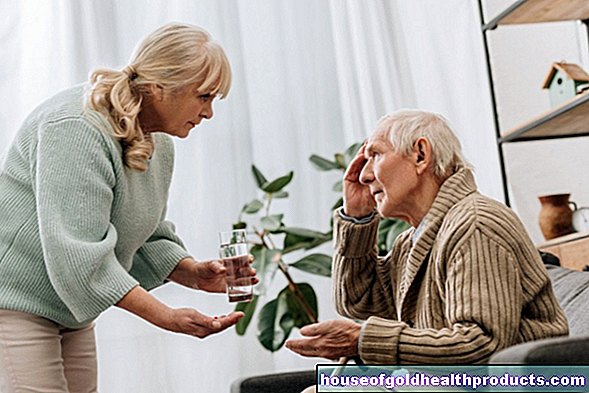
Pain therapy
Scoliosis pain in the back, neck or shoulders, but also headaches, are usually treated with painkillers in tablet form. Sometimes plasters that give off heat also help.Local anesthetic injections in the back are only used for severe pain. They arise in the context of scoliosis, for example, due to wear damage to the spine and constricted spinal cord nerves.
The so-called transcutaneous electrical nerve stimulation (TENS) sometimes helps against pain in scoliosis. Electrodes are applied to the skin over the painful area. These electrodes release electrical impulses that act on underlying nerves. They thus inhibit the transmission of pain from these nerves to the brain. The German Scoliosis Network also lists acupuncture as part of a comprehensive scoliosis therapy - it too is supposed to relieve pain in some patients.
Scoliosis: exercises
In the case of slight curvatures of the spine, physiotherapy exercises are suitable as scoliosis therapy. They are supposed to correct the posture. In addition to physiotherapy applications, there are also exercises for scoliosis that the patient can perform at home. Exercises are intended as part of scoliosis therapy
- improve posture
- strengthen the muscles
- Eliminate front and back curvature
- Increase lung and heart function
There are now many methods of treating scoliosis using exercises.
Read everything you need to know about scoliosis exercises here.
Scoliosis: disease course and prognosis
The course of scoliosis is very different. In principle, the earlier a spinal curvature is detected, the more likely it will progress.
Infant scoliosis is an exception. Within the first two years of life, a crooked spine regresses independently in up to 96 percent of cases. In addition, it can be positively influenced by suitable positioning measures and physiotherapy.
If a residual scoliosis of over 20 degrees remains, the parents of the affected baby must expect the scoliosis to progress.
Risk of worsening scoliosis
If scoliosis only occurs in the following years of life, the prognosis depends on various criteria. For example, underlying diseases of the muscle or nervous system can worsen the course of the disease. In idiopathic scoliosis, other factors are important in addition to age (possible residual growth):
- Exit Cobb angle
- Risser stage (skeletal maturity)
- Time of the first menstrual period (menarche, proven connection with relapsing bone growth in the following years)
The Cobb angle has the greatest importance at the beginning of the diagnosis. The guidelines indicate the likelihood of idiopathic scoliosis increasing, depending on the degree of curvature and age, as follows:
|
Cobb angle in degrees |
10-12 years |
13-15 years |
16 years |
|
less than 20 |
25 percent |
10 percent |
0 percent |
|
20-29 |
60 percent |
40 percent |
10 percent |
|
30-59 |
90 percent |
70 percent |
30 percent |
|
greater than 60 |
100 percent |
90 percent |
70 percent |
Course of the disease in old age
Scoliosis can worsen in adulthood. This is especially true if the Cobb angle is over 50 degrees at the end of the growth. Calculations on thoracic and lumbar scolioses have shown that the curvature increases by about 0.5 to one degree annually.
With severe scoliosis, especially in the lower back, the risk of painful complaints increases. Particularly pronounced curvatures can also irritate the spinal nerves and cause abnormal sensations or pain.
Caution: If the scoliosis reaches a value of around 80 degrees, it can reduce life expectancy.
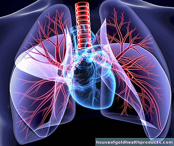
In the case of very pronounced courses, breathing becomes more and more difficult due to the increasing deformation. The chest is barely mobile (rigorous thorax) and the lung volume decreases. On the side of the curve, the lungs become overinflated (emphysema). The other half of the lung is hardly ventilated and the lung tissue collapses in places (atelectasis).
There is a risk of serious complications such as pneumonia, chronic bronchitis or inflammation of the lungs (pleurisy). In addition, the heart is also increasingly stressed (cor pulmonale).
Complications after scoliosis surgery
As with any surgical procedure, an operation on the spine also carries certain risks such as bleeding, infections (especially in acne patients) or wound healing disorders. Sensory disturbances or paralysis usually do not occur in idiopathic scoliosis. However, surgical scoliosis therapy can lead to nerve or spinal cord injuries.
However, the likelihood of such a complication is very low. According to studies, it is 0.3 to 2.5 percent. The risk increases if a major procedure is performed and other diseases (especially the spinal cord) exist. In some cases - for example with spinal cord diseases - the doctors let the person wake up during the operation and check their movements and sensations on the skin.
Effusions and tires
If the operation was carried out through the chest cavity, fluid may also accumulate in the chest. These are led out of the body through a tube (drainage). Under certain circumstances, a lung collapses, medical pneumothorax (short: Pneu). Here, too, a special drainage system is used so that the lungs can unfold again.
Correction loss
After some stiffening operations, the counter-curvature of a scoliosis can also intensify. In addition, the correction achieved is sometimes partially lost in the first few years after the procedure. Usually, however, scoliosis stabilizes after the operation.
Loss of correction can be problematic in young patients who are stiffened at an early age (crack 0). As the vertebral bodies continue to grow, the spinal torsion increases in many cases. Doctors speak of the so-called crankshaft phenomenon. To prevent this, stiffening scoliosis therapy is usually given from both the front and the back.
Other special complications include metal breaks in the rods and screws used in an operation. In these cases, there is almost always a loss of correction. In some stiffening operations, the vertebral bodies do not grow together as planned. "Wrong" joints, so-called pseudarthroses, are formed. They can cause persistent pain (especially in the case of lumbar coliosis).
In some patients, a straightening operation using rods (Harrington rods) develops a flat back. The naturally existing forward curvature of the lumbar spine is eliminated. As a result, the adjacent vertebrae and intervertebral discs wear out faster and cause painful discomfort.
Scoliosis and pregnancy
Contrary to popular belief, scoliosis does not have a negative impact on pregnancy. It does not matter whether the patients were treated conservatively (physiotherapy, corset) or surgically. As with all pregnant women, people with scoliosis can experience deeper back pain, but an increase in the Cobb angle has not yet been proven.
Check-ups
Depending on the extent of the scoliosis, the doctor will regularly check the curvature. Children's spinal curvatures below 20 degrees are checked approximately every three to six months through physical exams. If the doctor suspects an increase in curvature, he will order an X-ray. Scolioses over 20 degrees are checked at least once a year by means of an X-ray examination. Clinical examinations are also carried out at least every six months as part of scoliosis therapy.
If the growth is complete and the Cobb angle is less than 20 degrees, no further control is required. In the case of scoliosis of 20 to 40 degrees that has not been operated on, the doctor will carry out a check-up about two to four years after the growth has finished. If the curvature has increased by five degrees, further checks follow. If adults suffer from scoliosis over 40 degrees, the guidelines recommend an annual review.
If the affected person has been operated on, no further routine examinations are necessary two years after the operation with stable stiffening and a Cobb angle of less than 40 degrees.
Living with Scoliosis
In most cases, patients can live well with their scoliosis. It is important to work actively against the spinal deformity. Incorporate scoliosis exercises into your everyday life. Do (school) sports. If you have concerns about some activities, you should definitely consult your doctor.
If your scoliosis affects you in everyday life, for example at work or in your free time, do not hesitate to ask for help. Contact your employer, physiotherapist, or friends. Some of those affected also get involved in self-help groups. If scoliosis is a burden on your psyche or that of your child, psychotherapeutic treatment can also be useful. By being open and active, you can collect valuable tips and prevent the progression of your scoliosis.
Additional information:
Guidelines:
- Guidelines for the special rehabilitation concept for spinal deformities of the Rehabilitation and Physical Medicine Section of the DGOOC by orthopedists for orthopedists (as of 2012)
- Guidelines on idiopathic scoliosis in growing age (status: 2009, in revision)