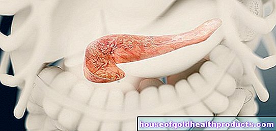
Aortic valve
Eva Rudolf-Müller is a freelance writer in the medical team. She studied human medicine and newspaper sciences and has repeatedly worked in both areas - as a doctor in the clinic, as a reviewer, and as a medical journalist for various specialist journals. She is currently working in online journalism, where a wide range of medicine is offered to everyone.
More about the experts All content is checked by medical journalists.The aortic valve is one of the four heart valves. It sits between the left ventricle and the main artery (aorta). There it acts as an outlet valve: it only allows blood to flow in the direction of the aorta. If the heart valve is narrowed or does not close tightly, it interferes with blood flow. Read everything you need to know about the aortic valve!
Aortic valve: pocket valve in the left heart
The aortic valve acts as a valve between the left heart chamber (ventricle) and the outgoing main artery (aorta). In terms of construction, it is a so-called pocket flap: It consists of three crescent-shaped "pockets", the shape of which is reminiscent of swallow nests. Due to their position and shape, they are called the rear, right and left semilunar valves and consist of a double layer of the endocardium (inner wall of the heart). Like the other valves, the aortic valve is attached to a fibrous ring of the heart skeleton.
Function as an exhaust valve
The aortic valve opens in the direction of the main artery (aorta) when, during systole (ventricular contraction), blood is pumped from the left ventricle into the aorta and thus into the large bloodstream. As long as the pressure in the left ventricle exceeds that in the aorta (which is the case with systole), no blood can flow back into the chamber. If, however, the ventricle relaxes during the following diastole (relaxation of the ventricle) in order to take in the blood from the left atrium, the pressure in the ventricle drops compared to that in the aorta. The blood could flow back; but the aortic valve prevents this backflow:
The flat pockets of the valve fill with blood, which is caught in them when the backflow begins. This puts the edges of the pockets close together. In addition, the pockets usually have a small nodule in the middle of their free edge. When the blood flows back against each other, these nodules get caught and thus additionally seal the center of the aortic valve. This securely closes the connection between the left ventricle and the aorta.
The doctor can perceive the closure of the aortic valve with the stethoscope as a second heart sound.
Common aortic valve problems
Aortic valve stenosis (aortic stenosis) is what doctors call a narrowed aortic valve. It is usually acquired, more rarely congenital. The most common cause of aortic stenosis are degenerative changes due to arteriosclerosis: deposits of calcium in the heart valve impair its mobility. It is now more difficult to pump the blood out of the left ventricle, and the pressure in the ventricle increases. As a result, the chamber wall thickens (hypertrophy).
In the case of aortic valve insufficiency, the heart valve no longer closes tightly, so that blood flows back from the aorta into the left ventricle during diastole. The now larger amount of blood loads the left ventricle (volume load), which eventually expands (dilation). With aortic regurgitation, the heart wall can also thicken.
People whose aortic valve consists of only two pockets are particularly susceptible to these diseases. This so-called bicuspid (double-leaved) aortic valve is the most common congenital heart valve defect. It occurs in about one to two percent of the population and mainly men.
Aortic valve disorders are best heard by the doctor with a stethoscope to the right of the sternum, roughly between the second and third ribs.
In both stenosis and insufficiency, it may be necessary to replace the diseased aortic valve with a prosthesis.
Tags: pregnancy organ systems interview


-mit-mickymaus-am-tannenbaum.jpg)