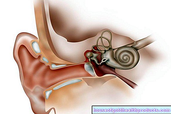
Eye muscles
Eva Rudolf-Müller is a freelance writer in the medical team. She studied human medicine and newspaper sciences and has repeatedly worked in both areas - as a doctor in the clinic, as a reviewer, and as a medical journalist for various specialist journals. She is currently working in online journalism, where a wide range of medicine is offered to everyone.
More about the experts All content is checked by medical journalists.The eye muscles allow the eyeball to move. Optimal vision would not be possible without precise eye movements. The eye muscles help to scan the environment with fast, synchronous eye movements and to change the curvature of the eye lens in order to enable near and far vision. Read more about the eye muscles!
What are the eye muscles?
Six eye muscles move the human eye in all directions. There are four straight and two oblique eye muscles.
Eye muscles straight
The four straight eye muscles are flat, thin muscles about an inch wide. They pull from the upper, lower, middle and outer walls of the orbit (eye socket) to the edge of the cornea. The optic nerve runs in the space behind the eyeball, which the eye muscles enclose in a pyramidal shape.
The four straight eye muscles pull the eye in the following directions:
- upwards and slightly inwards (rectus superior muscle)
- downwards and slightly inwards (rectus inferior muscle)
- towards the middle - i.e. towards the nose - (musculus rectus medialis, the strongest of the eye muscles)
- outward (rectus lateralis muscle)
Oblique muscles
The two oblique eye muscles pull medially from the front (towards the center of the face) to the rear laterally (towards the outside). They ensure the following eye movements:
- Pull outwards and twist downwards inwards (Musculus obliquus superior)
- Pull outwards and twist upwards outwards (Musculus obliquus inferior)
Ciliary muscle
Another eye muscle is the ciliary muscle, but it is not involved in the movement of the eye. Instead, the function of the ciliary muscle is to accommodate the eye:
The ciliary muscle is part of the ciliary body (radiating body) - the annular middle layer of the eyeball. Processes extend from the ciliary body to the lens of the eye, between which the lens hanging strap is stretched.
- When the ciliary muscle tenses, the hanging ligament slackens and the lens arches more strongly, following its own elasticity. This will bring the near area into focus.
- In the opposite case, i.e. when the ciliary muscle relaxes, the lens becomes flatter and thus the vision in the distance is focused.
What is the function of the eye muscles?
The eye muscle function is to move the eyeball. A sharp image of our environment can only be created in a small area of the retina, the point of central vision (fovea). At a distance of one meter we can see clearly an area with a diameter of only nine centimeters.
In order to still be able to perceive everything around us clearly, the eye must be able to scan every image that penetrates the eye from the outside with rapid movements. These leaps in view are called saccades. The eye is repeatedly directed from the rest position at high speed to the next target. So we do not capture our entire field of vision at once, but “little by little”.
When reading, for example, these saccades occur at intervals of 200 to 400 milliseconds.
In contrast to the saccade, which is necessary to capture a stationary image, moving objects are perceived by the eyes making a subsequent movement without jerking. This movement is much slower than the jerky saccades.
Both eyes must be moved absolutely synchronously to avoid double vision. The eye must also compensate for head or body movements through movement in order to avoid blurring on the retina. The eye muscles make this possible.
What problems can the eye muscles cause?
If the eye muscles do not work absolutely synchronously, i.e. there is a deviation of the eye axes, then it comes to squint (strabismus). The squint angle is always the same. This squint almost always begins in childhood.
Squinting also occurs when one eye muscle is paralyzed. The squint angle then changes with the eye movement and is greatest when looking in the direction in which the main effect of the paralyzed muscle lies. As a result, double images emerge, which the person concerned tries to compensate for with the posture of his head.
A paralysis of the eye muscles can result from diseases of the eye socket (orbit) or from paralysis of the eye muscle nerves.
Tags: skin care book tip toadstool poison plants


.jpg)