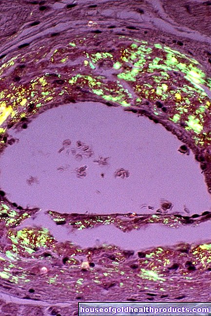
Renal pelvis
Eva Rudolf-Müller is a freelance writer in the medical team. She studied human medicine and newspaper sciences and has repeatedly worked in both areas - as a doctor in the clinic, as a reviewer, and as a medical journalist for various specialist journals. She is currently working in online journalism, where a wide range of medicine is offered to everyone.
More about the experts All content is checked by medical journalists.The renal pelvis (pelvis renalis) is part of the urinary tract and acts as a collecting basin for the newly formed urine. A funnel-shaped (dendritic) to round (ampullary) hollow kidney system, into which the collecting tubes guide the urinary tract, arises from the unifying kidney calyxes. Read everything you need to know about the kidney pelvis!
What is the renal pelvis?
The renal pelvis is a sack that is flat at the front (towards the abdomen) and back (towards the back) within the kidney. The calyxes, which have absorbed the urine produced, pass it on to the renal pelvis, where it is collected like a small collecting funnel before it flows over the ureter towards the bladder.
There are two different anatomical variants: the calyxes can release urine directly into a large pelvis - this is called the ampullary type - or they form intermediate pieces of different lengths, which then form a narrow, tubular renal pelvis - this is called the dendritic type.
The renal pelvis is lined with a typical epithelial layer (urothelium) - the same tissue as the entire lower urinary tract. This type of tissue is characterized by so-called transitional cells, which - when there is little urine - have a tall shape, but flatten out as the urinary tract becomes more full. So you can easily adapt to different filling states. An additional expansion of the lumen is possible through folds in the cell membrane.
What is the function of the renal pelvis?
The renal pelvis directs the urine supplied via the calyxes into the ureters, from where the urine enters the urinary bladder and then outwards via the urethra. Like the upstream kidney calyxes and the downstream ureters, it is equipped with smooth muscles in its wall, which ensure that the urine is transported.
Where is the renal pelvis located?
The renal pelvis is located in the back of the sinus, facing the back - the area on the middle edge of the kidney where there is a deep pit. There the renal artery enters the organ and the renal vein and ureter (ureter) lead out.
The renal pelvis descends slightly downwards from the center of the kidney. This position can be used well by the surgical side if operations (for example the removal of kidney stones) are necessary - this area can be easily reached from the back.
What problems can the renal pelvis cause?
A bacterial infection can cause inflammation of the kidneys (pyelitis), which in most cases is also accompanied by inflammation of the kidney as a whole (pyelonephritis).
Renal pelvis stones (nephrolithiasis) are urinary stones that more or less completely fill the renal pelvis. Large stones in the ureter can cause the pressure to rise to such an extent that a tear occurs at the junction with the kidney parenchyma (fornix rupture).
An enlargement (pyelectasia) that does not involve the calyx system can be congenital or develop after infection. A lack of the mutual kidney or tumors can also lead to this.
Hydronephrosis is a kidney congestion in which the renal hollow system expands due to an obstruction of the drainage with subsequent backflow. As a result, kidney tissue dies.
A renal pelvic fistula results either from an injury or from the breakthrough of a tumor - but it can also be created for therapeutic reasons. This is a tubular connection between the renal pelvis and either the surface of the skin or another hollow organ such as the urinary bladder.
Last but not least, there are also tumors in this area, such as renal pelvic carcinoma or benign renal pelvic papilloma.
Tags: gpp toadstool poison plants interview


.jpg)

