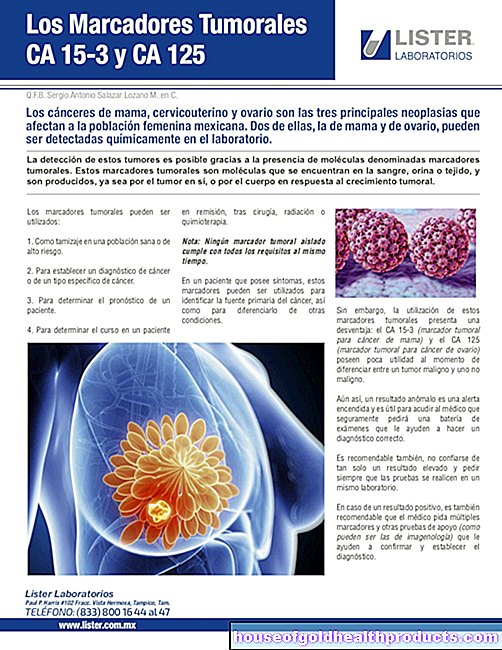
Angiography
Valeria Dahm is a freelance writer in the medical department. She studied medicine at the Technical University of Munich. It is particularly important to her to give the curious reader an insight into the exciting subject area of medicine and at the same time to maintain the content.
More about the experts All content is checked by medical journalists.In angiography, vessels are visualized with the help of diagnostic imaging methods such as x-rays, magnetic resonance imaging (MR angiography) or computed tomography (CT angiography). The doctor can thus diagnose and assess vascular diseases. Read all about angiography, how it is done and the risks it carries.

What is an angiography?
Angiography is a radiological examination in which the vessels are filled with contrast medium and made visible with the help of x-rays, magnetic resonance tomography or computed tomography and shown on the so-called angiogram. Depending on the type of vessels, a distinction is made between the angiography of the arteries (arteriography), the veins (venography) and the lymphatic drainage pathways (lymphography).
When do you do an angiography?
Angiography is used to diagnose diseases that are associated with narrowing or occlusion of the vessels.
Angiography: heart
Heart angiography is also known as coronary angiography. It makes the coronary arteries visible, which can be changed or blocked in the course of coronary heart disease or a heart attack. In addition, the interior of the heart can be displayed and their size and function assessed.
Angiography: eye
With the help of so-called fluorescence angiography, the doctor can diagnose age-dependent macular degeneration (disorder of the retina) by assessing the fine blood vessels in the retina. A special green dye (fluorescein) is used instead of the contrast agent.
Angiography: brain
With cerebral angiography (Latin: cerebrum = brain), both the blood vessels in the brain and the supplying vessels in the neck area can be visualized if there is a suspicion of brain tumors, cerebral haemorrhage or vascular diseases.
Angiography: legs
The arteriography of the leg and pelvic vessels is used to detect vascular constrictions, for example in diabetics. The venography is carried out if thrombosis is suspected and for therapy planning in the case of varicose veins.
If there is a contrast agent intolerance, CO2 angiography can be performed on the legs, in which the contrast agent is replaced by carbon dioxide.
What do you do with an angiography?
Before the actual examination, your doctor will take a medical history and explain the risks and benefits of the procedure. In addition, the blood values are measured and tested for a possible contrast agent allergy.
With conventional angiography, a thin plastic tube (catheter) is first inserted into the artery, vein or lymphatic vessel, usually under local anesthesia, and pushed forward until shortly before the vessel section to be examined. After the injection of the contrast medium, which fills the vessels, the corresponding area of the body is x-rayed. The contrast agent appears white on the X-ray because it absorbs the X-rays. This means that the interior of the vessels can also be seen on the angiogram. Finally, the catheter is removed and a pressure bandage is applied over the puncture site.
A special form is digital subtraction angiography, in which recordings are made before and after the contrast agent is distributed. A computer removes identical areas from both images, making the changes in the contrast medium-filled vessels particularly visible.
In contrast, in CT angiography and MR angiography, the contrast agent does not have to be injected directly into the vessel to be visualized, but is usually administered via an arm vein or artery. No contrast agent is required for time-of-flight MR angiography (TOF angiography), since the images are created by magnetizing freshly flowing blood.
What are the risks of angiography?
Angiography is a relatively uncomplicated examination. When the contrast agent is injected, the mouth may feel warm or taste unpleasant. These harmless side effects disappear immediately after the injection.
In rare cases, people are hypersensitive to the contrast agent or develop an allergic reaction. The allergy, renal insufficiency or hypothyroidism must be clarified before the examination, since in these cases no contrast agent should be given.
A vascular puncture can lead to bleeding, bruising, thrombosis (clots), embolism (vascular blockage due to thrombosis), vascular injuries or infections.
What do I have to consider after an angiography?
After the angiography, you should take it easy and drink as much as possible so that the contrast agent is eliminated quickly. You should also avoid heavy physical labor in the following days. If you suddenly experience dizziness, headache, nausea or a racing heart, please notify a doctor immediately.
Tags: gpp teenager hair