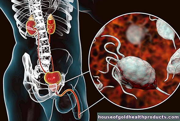
Scintigraphy
Dr. med. Philipp Nicol is a freelance writer for the medical editorial team.
More about the experts All content is checked by medical journalists.Scintigraphy is a nuclear medicine procedure for displaying body tissue. In doing so, one uses weakly radioactive substances that accumulate in various organs. The radiation emitted by them is measured and provides information on the metabolic activity and blood flow in the tissue.Here you can read everything you need to know about the different types of scintigraphy, when to use them and what risks they involve.

What is a scintigraphy?
Scintigraphy is an examination method from the field of nuclear medicine: the patient is injected with low-level radioactive substances as drugs for diagnostic purposes. There are two types of these so-called radiopharmaceuticals:
Some radioactive substances are administered directly. An example of such radionuclides is radioactive iodine, which mainly migrates to the thyroid.
In other cases, the radioactive substance is bound to a carrier (such as certain proteins or salts) that is largely or exclusively metabolized in certain organs. The radioactive marking of such a carrier is called a tracer.
In the target tissue, the radiopharmaceutical accumulates particularly in areas with high metabolic activity and good blood circulation. It disintegrates, giving off so-called gamma rays, which are measured by a special camera (gamma camera). A computer uses this to calculate an image of the examined body region (scintigram).
For example, scintigraphy can be used to precisely localize sources of inflammation in the body. The metabolism runs faster in an inflamed area. An increased metabolic activity can also speak for a tumor, and a reduced activity can be an indication of dead tissue.
Scintigraphy can be used to examine a large number of different tissues, for example bones, the thyroid gland or the heart muscle.
Further information: bone scintigraphy
The method is particularly suitable for examining bones. You can read more about this in the article on bone scintigraphy.
Further information: Thyroid scintigraphy
You can find out which useful information an examination of the thyroid gland provides in the article Thyroid scintigraphy.
Further information: Myocardial scintigraphy
With the myocardial scintigraphy, the condition of the heart muscle (myocardium) can be checked. Read more about this in the article Myocardial Scintigraphy.
Somatostatin receptor scintigraphy (octreotide scintigraphy)
This special form of scintigraphy can be used to detect certain tumors that can hardly be diagnosed with other methods (X-ray, ultrasound, CT or MRT). These so-called neuroendocrine tumors (NET) are often located in the abdomen (intestines, stomach, pancreas) and produce hormones. On its surface there are docking points for the hormone somastatin (somatostatin receptors). The tumors can be made visible through the binding of suitable radioactively labeled substances (such as octreotide) to this receptor.
SPECT and SPECT / CT
SPECT (Single, Photon Emission Computed Tomography) is a further development of the procedure in which several gamma cameras move around the patient. In contrast to normal “planar” scintigraphy, three-dimensional sectional images can be generated.
There are now also devices that can be used to perform SPECT in combination with computed tomography (CT) (SPECT / CT). The metabolically active regions in the body that are imaged using SPECT can be displayed more sharply thanks to CT.
When do you perform a scintigraphy?
In contrast to other imaging methods such as computed tomography (CT) or magnetic resonance tomography (magnetic resonance tomography, MRT), scintigraphy provides information about the activity of tissue. Since tumors often have increased metabolic activity, scintigraphy is used particularly frequently in cancer medicine. There are also other possible uses for the nuclear medicine procedure, such as:
- Clarification of suspicious lumps or an overactive thyroid
- Examination of kidney function (e.g. if renal artery stenosis is suspected)
- Examination of the blood flow and ventilation of the lungs if a pulmonary embolism is suspected (perfusion ventilation scintigraphy of the lungs)
- Clarification of diseases or injuries to bones (such as infections, osteonecrosis, osteoporosis, tumors, fractures)
- Function test of the heart muscle (for example after a heart attack or coronary heart disease)
In pregnant and breastfeeding women, a scintigraphy should only be carried out in absolutely exceptional cases, if the information gain to be expected from the examination is greater than the health risk from the radiation exposure.
What do you do with a scintigraphy?
The scintigraphy is performed by a specialized doctor, a nuclear medicine specialist. He or she will have a detailed discussion with you before the examination. He will inform you about the advantages and risks of the examination and ask you about previous illnesses and regular medication intake.
Special preparation before the scintigraphy (such as being sober) is usually not necessary. For the examination, the radioactive substance will be injected into you through a vein. You may then have to wait a certain amount of time (a few minutes to hours) for the radioactive substance to reach the target organ. The actual recordings usually only take a few minutes.
The examination itself is completely painless. In contrast to CT or MRT examinations, you do not have to go into a "tube" for normal scintigraphy, as the gamma camera can be moved freely.
What are the risks of scintigraphy?
Side effects with scintigraphy are very rare. The administered radiopharmaceutical may cause a temporary sensation of heat, skin reactions (itching, redness, etc.), a metallic taste in the mouth, or slight nausea. Patients should speak to their doctor about possible scintigraphic side effects after the examination.
In the long term, radiation exposure poses a certain health risk. However, the radiation exposure is low (comparable to that of an X-ray). In addition, the body quickly excretes the substance again. How high the health risk from radiation is depends primarily on the type and amount of radioactive material used and the region of the body examined.
What do I have to consider after a scintigraphy?
After a scintigraphy, it is important that the body excrete the radioactive material quickly in order to reduce the radiation exposure. The radionuclide is mainly excreted through the kidneys. Therefore, after the scintigraphy, you should drink a lot and go to the toilet frequently. If you are only allowed to drink a certain amount of fluids per day because of a kidney or heart failure, the attending physician will provide you with further information.
Immediately after the scintigraphy, you will emit light radioactive radiation. You should therefore avoid close contact with pregnant women, breastfeeding women and young children for a few hours.
Tags: toadstool poison plants alternative medicine Menstruation