Cataract
and Martina Feichter, medical editor and biologist and Carola Felchner, science journalistSophie Matzik is a freelance writer for the medical team.
More about the expertsMartina Feichter studied biology with an elective subject pharmacy in Innsbruck and also immersed herself in the world of medicinal plants. From there it was not far to other medical topics that still captivate her to this day. She trained as a journalist at the Axel Springer Academy in Hamburg and has been working for since 2007 - first as an editor and since 2012 as a freelance writer.
More about the experts
Carola Felchner is a freelance writer in the medical department and a certified training and nutrition advisor. She worked for various specialist magazines and online portals before becoming a freelance journalist in 2015. Before starting her internship, she studied translation and interpreting in Kempten and Munich.
More about the experts All content is checked by medical journalists.Cataracts are an eye disease in which the otherwise clear lens becomes increasingly cloudy. As a result, eyesight steadily decreases. Cataracts are often age-related. However, it can also be the result of metabolic diseases or eye malformations and injuries, for example. Cataracts can be treated well with surgery. If left untreated, it can lead to blindness. Read more about the symptoms, causes and treatment of cataracts here!
ICD codes for this disease: ICD codes are internationally recognized codes for medical diagnoses. They can be found, for example, in doctor's letters or on certificates of incapacity for work. H28H25Q12H26
Brief overview
- Symptoms: increasing deterioration of eyesight, sensitivity to glare, seeing "like through a veil / fog"
- Causes: mostly aging processes of the eye, sometimes other diseases (e.g. diabetes mellitus, eye inflammation), eye injuries, congenital eye malformations, exposure to radiation, heavy smoking, medication
- Diagnostics: i.a. Discussion with the patient, various eye examinations (e.g. using a slit lamp), possibly further examination if an underlying disease is suspected (such as diabetes)
- Treatment: surgery
- Prognosis: generally good chances of success with surgery
Cataracts: symptoms
If the view becomes cloudy and the world seems to disappear behind a veil, this could be a sign of the eye disease cataracts. "Gray" because the lens turns greyish in advanced disease and thus becomes cloudy. The name suffix "Star" is derived from the stare that sufferers have when they are (almost) blinded by the eye disease.
The medical term for cataracts - cataract - comes from the Greek and means "waterfall". It used to be assumed that coagulated liquid in the eye caused the lens to become cloudy.
-
Cataracts: "Once a year for an eye check"
Three questions for
Dr. med. habil. Wolfgang Herrmann,
Specialist in ophthalmology -
1
Does everyone get (age-related) cataracts?
Dr. med. habil. Wolfgang HerrmannIncreasing clouding of the lens is part of aging. We ophthalmologists only speak of a "cataract" when the cloudy lens reduces visual acuity and the quality of vision deteriorates. Conditions like diabetes, an unhealthy diet, or smoking can cause the lens to cloud more quickly. UV light also favors cataracts - sunglasses can protect here.
-
2
Can I test myself whether I have cataracts?
Dr. med. habil. Wolfgang HerrmannThose affected often notice that they have less vision or that they are severely blinded when driving at night. Colors are also perceived differently. These are all vision problems that can also be caused by other eye diseases. That is why you should see an ophthalmologist. Only he can unequivocally recognize a cataract. My advice: Older people should have their eyes checked once a year.
-
3
How quickly do you have to operate?
Dr. med. habil. Wolfgang HerrmannIn general, cataracts are operated on if you have problems in everyday life or if you have too bad eyesight to be able to drive. But don't worry: with over 700,000 procedures a year, cataract treatment is one of the most common operations. And because the methods continue to improve - you can also correct ametropia - most of them can be operated on sooner rather than later.
-
Dr. med. habil. Wolfgang Herrmann,
Specialist in ophthalmologyHerrmann heads the eye clinic at the Hospital of the Brothers of Mercy in Regensburg. His main focus is on refractive surgery for ametropia and the treatment of retinal diseases.

Cataracts: symptoms in the course of the disease
Cataracts cause different symptoms depending on the stage. At the onset of the disease, vision deteriorates and those affected become increasingly sensitive to glare. In the middle of the field of vision, a kind of fog arises through which objects are perceived as blurred or as if they are behind a veil.
This fog becomes more and more dense over time and spreads over the entire field of vision in the course of the disease. Colors, contrasts and contours gradually fade and seem to merge with one another. Spatial perception and thus also the ability to orientate deteriorate.
This is how the picture changes with cataracts
Individual and complete failures of the field of vision, as they occur in cataracts (glaucoma), do not occur in cataracts.
In the further course of the disease, symptoms of cataracts appear, which those affected can stress in everyday life. This includes:
- significant sensitivity to glare (e.g. in bright sunlight or flash light)
- indistinct visual perception
- poorer light-dark adaptation
- Exertion while reading or watching TV
- limited spatial vision
- Road traffic uncertainty
These symptoms can vary in severity in each patient. They do not necessarily have to occur (all).
Late-stage cataracts make normal everyday life almost impossible: visual performance can deteriorate so dramatically within a short period of time that it amounts to blindness.
Cataracts: Symptoms often not recognized or misinterpreted for a long time
If cataracts affect both eyes, it is often difficult for those affected to assess how far the deterioration in vision is. This becomes dangerous in road traffic, for example.
Another problem is that many people with cataracts initially ignore, cover up, or attribute the symptoms to other causes such as fatigue. Especially in cataracts, which arise as a result of the natural aging process, the symptoms are often justified with the age-related deterioration of the eyes - and not with a manifest eye disease such as cataracts.
Cataracts: Family members should watch out for signs
Precisely because those affected often incorrectly assess or deny the deterioration in vision, it is important that the relatives know the symptoms of cataracts and interpret them correctly. In the early stages of the disease, people become less confident about normal activities, such as driving a car or reading. This can be seen, for example, from the fact that the patients often show a strained expression on their faces during these activities.
In later stages, the deterioration in vision can become so severe that those affected often go wrong if something is offered to them or if they want to take something into their own hands. In addition, it takes them a long time to find their way around an environment that they are not familiar with. Therefore, they often avoid unknown places.
Orientation is also more difficult in your own home environment. Most people with cataracts tend to be meticulous in order to keep everything in order so that they can find everything without full eyesight.
Congenital cataracts: symptoms
Children can also develop cataracts. Doctors then speak of a child's or congenital cataract. The lens opacity can already exist at birth or develop in the course of the first few years of life. The first sign of this is often that the children are starting to squint (strabismus).
Parents should not ignore this, but take it seriously. If left untreated, the loss of visual acuity can impair the development of the visual system, which reacts particularly sensitively to disorders in the first few months of life: If cataracts are not recognized and treated in the baby, it can lead to so-called weak vision (amblyopia).
This weak-sightedness can no longer be remedied by reaching puberty at the latest. You should therefore see a doctor immediately if the child shows signs of cataracts!
Cataracts: causes and risk factors
In the vast majority of cases, cataracts are age-related. But it can also have other causes such as metabolic diseases, other eye diseases or eye injuries. More on this below:
Natural aging process
The lens of the eye is usually clear and flexible so that the small muscles of the eye can deform it as needed. This deformation and the fluid that surrounds the lens enable us to see near and far objects equally sharply. This adaptation of the refractive power of the eye to objects at different distances is called accommodation.
This is how the eye is built
With increasing age, the flexibility of the lens of the eye naturally decreases, which can lead to lens opacity. That is why around 90 percent of all cataract cases are age-related cataracts. This gray age star occurs around the age of 60. According to statistics, almost half of 52 to 64 year olds have cataracts without knowing it. Because at the beginning of the disease, there is often no visual impairment perceptible. From the age of 65 almost everyone has a clouding of the eye lens.
Diabetes (diabetes mellitus)
In diabetes mellitus, the sugar content in the eye water (and blood) is increased. Excess sugar (glucose) accumulates in the lens, which causes it to swell. As a result, the arrangement of the lens fibers shifts and the lens becomes cloudy. Doctors speak of cataracta diabetica here.
In pregnant women with diabetes mellitus, the unborn child can develop cataracts.
Other metabolic diseases
In addition to diabetes, other metabolic disorders can also promote cataracts. These include, for example:
- Calcium deficiency (hypocalcaemia)
- Overactive parathyroid gland (hyperparathyroidism)
- Excess ferritin in the blood (ferritin is an iron storage protein)
- Galactosemia (a congenital disorder of using the sugar galactose in breast milk)
Eye diseases
Cataracts can also result from other eye diseases and are called cataracta complicata. Possible triggers are, for example, eye inflammation (such as inflammation of the middle skin of the eye = uveitis) or severe myopia.
Eye injuries
A bruise of the eyeball from a punch or a tennis ball can also cause cataracts, such as a stab wound or a deeply penetrated foreign body in the eye. Such injury-related cases of cataracts are summarized under the technical term cataracta traumatica.
Congenital malformations of the eye
If cataracts are congenital (cataracta congenita), there can be two reasons:
- Genetic defect: Around 25 percent of all congenital cataract diseases are based on a genetic defect that leads to malformation of the eye and thus to opacity of the lens.
- Infectious diseases during pregnancy: Certain infections in pregnant women (rubella, toxoplasmosis, herpes) can lead to the child being born with a cataract.
Other causes
Lens metabolism defects, malnutrition, heavy smoking, radioactive radiation and ultraviolet light (UV light) can also trigger cataracts. Drugs or poisoning are very rarely the cause of the clouding of the lens.
Cataracts: examinations and diagnosis
A detailed examination by the ophthalmologist is necessary for the diagnosis of cataracts.
anamnese
It starts with a detailed conversation between doctor and patient to collect the medical history (anamnesis): Among other things, the doctor asks about the exact symptoms and any existing underlying diseases (diabetes, eye diseases, etc.).
Eye exams
This is followed by various eye exams. To do this, the pupil is sometimes first dilated with the help of special eye drops. The following tests help diagnose cataracts:
- Brückner test: In this examination, the doctor x-rays the eye. Since the retina reflects some of the light, lens opacities can be seen as dark spots.
- Slit lamp examination: The slit lamp is a microscope with a light source that can be swiveled to both sides. The bundled, slit-shaped light beam penetrates the transparent eye area. In this way, the doctor can also examine the retina at the back of the eye and see what type of cataract is present and what could be the cause.
- Corneal examinations: The doctor can measure the thickness of the cornea (pachymetry) and map its top and back surface using computer-aided methods (Pentacam). The latter shows whether the cornea is evenly curved and whether the cell layer that supplies the cornea and ensures its transparency is in order (determination of the endothelial cell density).
- General eye test: The ophthalmologist also routinely checks general vision, for example using eye charts, and whether there are other eye diseases.
If cataracts are already well advanced, the lens opacity can already be seen with the naked eye.
Other investigations
Sometimes cataracts are the first sign of another underlying disease. Additional examinations, such as skin and muscle tests and blood tests, are therefore necessary, especially in young patients. For example, tetany (pathological muscle spasms), myotonia (muscle disease), skin diseases, Wilson's disease (congenital disorder of the copper metabolism) or diabetes mellitus can be determined.
Cataracts: treatment
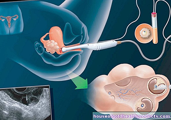
The only effective way to treat cataracts is through surgery (cataract surgery). The cloudy lens is removed and replaced with an artificial lens. As a rule, nowadays the surgeon no longer removes the entire lens, but leaves the lateral and rear eye capsules in the eye.
The cataract operation is the most common operation on the eye. In Germany alone, the operation is performed around 700,000 times a year, and more than 100 million times worldwide.
The procedure is a so-called microsurgical operation, i.e. it is carried out with a surgical microscope. This is possible both in the hospital and in an ophthalmologist's practice. The artificial lens used remains in the eye for life, so it does not have to be replaced after a while.
Cataract surgery: when is it necessary?
When a cataract is operated on depends on various factors. The doctor and patient jointly determined the time of the operation.
The subjective perception of visual impairment plays a role in the decision. If a person affected by cataracts feels severely impaired in everyday life and in their professional life, this speaks in favor of an operation.
In other cases, an objective deterioration in vision makes the procedure advisable or necessary: For example, people who drive a car have to have an eye test carried out at regular intervals. If you have a certain impairment of your eyesight, you can no longer drive - a good argument in favor of an operation.
In some professions, a certain visual performance is even a mandatory requirement, for example for pilots and professional drivers. Then cataract surgery is often necessary at an early stage of the disease. The subjective perception of the visual performance does not play a role here.
If possible, when deciding for or against an operation, the patient's fears regarding the operation on the eye are taken into account. If, however, the visual performance of an advanced cataract has deteriorated and even threatens to go blind, an operation should be carried out despite such fears.
Congenital cataracts should be operated upon immediately after diagnosis. Only then does the child have the chance to learn to see properly.
Lenses used
The intraocular lens used in cataract surgery is made of a plastic. It must have exactly the same refractive power as the removed body's own lens. The doctor calculates the appropriate lens power before the operation by measuring the patient's eye length with an ultrasound device and determining the refractive power of the cornea.
The artificial lenses used differ in terms of implantation location, material and their optical principles.
Differences in the place of implantation
Depending on the implantation site, a distinction is made between anterior chamber lenses, posterior chamber lenses and iris-borne lenses.
- Anterior chamber lenses (VKL) are inserted into the anterior chamber of the eye (in front of the iris) and attached there with two brackets in the angle of the chamber. They are only used for intracapsular cataract extraction (see below) - but only rarely because anterior chamber lenses can cause glaucoma or clouding of the cornea.
- Posterior chamber lenses (HKL) are inserted into their own capsular bag, which is located behind the iris. If, as with intracapsular cataract extraction, there is no longer a capsular bag, the lens is attached to the iris or sclera of the eye with two sutures.
- Iris-worn lenses (iris clip lenses) are attached to the iris with small brackets. Since this often injures the cornea, such lenses are no longer used in Germany. In many cases, already implanted iris-borne lenses are replaced by posterior chamber lenses.
Differences in lens material
In cataract surgery with a small incision, intraocular lenses made of silicone or acrylic are used, as these lens materials are foldable. These artificial lenses are inserted into the capsule in the folded state, where they then unfold by themselves. They are used exclusively as posterior chamber lenses.
An acrylic lens has a higher refractive index than a silicone lens and is therefore a little thinner.
Dimensionally stable lenses made of polymethyl methacrylate (PMMA, Plexiglas) can be used both as anterior chamber lenses and as posterior chamber lenses. A slightly larger incision is required for the implantation.
Differences in the optical principles
The optical principle is understood to be the properties of the lens that create the new “eyesight” in the person affected. Doctors differentiate between monofocal lenses and multifocal lenses.
- Monofocal lens: like normal glasses, they only have one focal point. It enables sharp vision either in the distance or in the vicinity. Before the operation, the patient has to decide whether he would prefer to live without "distance glasses" after the operation, but with reading glasses, or vice versa. The appropriate power of the artificial lenses is selected accordingly.
- Multifocal lens: It provides good visual acuity both far and near. Patients no longer need glasses for over 80 percent of their daily tasks. But multifocal lenses have two disadvantages: Contrasts are seen less sharply and the eye becomes more sensitive to glare.
Surgical methods
There are various methods of lens implantation to remove lens opacification. Which one is used in each individual case depends on the individual requirements and the stage of the disease.
Intracapsular Cataract Extraction (ICCE)
In this type of cataract surgery, the lens, including the capsule, is removed from the eye. This requires a relatively large incision (eight to ten millimeters) through the cornea. After that, the lens is frozen with a special cold stick and removed from the eye. The surgeon then inserts the artificial lens either into the anterior chamber (anterior chamber lens) or into the posterior chamber (posterior chamber lens) and mows the cut with a fine thread.
The intracapsular cataract extraction is usually only necessary at an advanced stage of the disease.
Extracapsular Cataract Extraction (ECCE)
With extracapsular cataract extraction, the surgeon opens the anterior lens capsule with an incision about seven millimeters long and removes the lens nucleus without comminuting it. The artificial lens is now inserted into the capsule that has remained intact.
This surgical method is gentle on the cornea. That is why it is mainly used when a well-advanced cataract has already damaged the thin, innermost layer of the cornea (corneal endothelium).
Phacoemulsification (Phaco)
During phacoemulsification, the cornea is opened with an incision about 3.5 millimeters wide. Then the doctor dissolves the lens nucleus with the help of ultrasound or a laser and sucks it off. The artificial replacement lens is now inserted into the intact cover of the lens (capsular bag): it is folded and pushed through the tiny opening and unfolds in the capsular bag itself. Two semicircular elastic straps on the edge of the lens ensure a secure hold in the capsular bag.
The tiny incision in phacoemulsification closes by itself after the operation without scarring. Finally, the surgeon only has to close the conjunctiva that was previously pushed back. Thanks to the small incision, with this surgical method it is possible to fit new glasses earlier than with the others and to resume normal everyday life.
The cataract operation
Cataracts usually appear on both sides. First, however, only one eye is operated on. As soon as this eye has healed, it is the turn of the second.
The procedure usually takes less than 30 minutes.
Outpatient procedure, local anesthesia
Cataract surgery is usually performed on an outpatient basis under local anesthesia. In most cases, the administration of suitable eye drops is sufficient for anesthesia. Alternatively, a local anesthetic can be injected into the skin next to the eye to be operated on. The entire eyeball becomes painless and can no longer be moved. The doctor can also give you a light sedative before the operation.
During the entire operation, your circulation will be monitored with the help of a blood pressure monitor, by measuring the oxygen saturation or with the help of an EKG.
After the operation, the operated eye is covered with an ointment bandage. You will need to remain in the hospital or doctor's office for some time to be monitored. If there are no complications, you can go home after a few hours. In the following period, regular check-ups by the attending physician are necessary.
What to watch out for after the procedure
Remember that you are not allowed to drive a car immediately after cataract surgery. So you should be picked up.
You can have light meals and drinks on the day of the operation. You can usually take your usual medication as usual, but you should discuss this with your doctor beforehand. This is especially advisable if you need medication for diabetes or blood thinning medication.
As long as the operated eye is covered with a bandage and the surgical wound has not yet healed, you should make sure that the eye does not come into contact with soap when showering and washing.
Physical exertion, swimming, diving, cycling and going to the sauna should be avoided in the initial period after the cataract operation. The same applies to activities that involve a lot of dirt or dust. You can usually read and watch TV again after a week.
You can usually have new glasses fitted four to six weeks after the cataract surgery. This does not make sense at an earlier point in time, as the eye has to get used to the new lens first.
If you notice the following symptoms some time after the cataract surgery, you should see an ophthalmologist:
- Deterioration in visual acuity
- increased redness of the eye
- Pain in the eye
Risks and Complications of the Surgery
About 97 to 99 percent of all cataract operations are without complications. However, as with any surgical procedure, there are also risks. These include:
Capsule tear
If the posterior capsule of the lens tears during the operation, complications can arise. The so-called vitreous body is located behind the lens of the eye.It consists of a gel-like, transparent mass and presses the retina, which is located in the back of the eye, against its surface. If the vitreous substance escapes through a crack in the lens, there is a risk of retinal detachment.
This risk exists in about six to eight percent of intracapsular operations; In an extracapsular operation, however, a capsule tear rarely occurs.
Bacterial infection
In an intracapsular cataract surgery, bacteria very rarely get inside the eye and cause inflammation (endophthalmitis). The affected eye can become blind as a result.
Bleeding
During cataract surgery, there may be an increase in pressure in the eye, which can burst blood vessels. Bleeding within the eye (intraocular) or within the capsule (intracapsular) is the result. However, they are very rare: such bleeding occurs in less than one percent of all cataract operations.
Corneal curvature
With the extracapsular surgical method, the incision causes a slightly greater curvature of the cornea than before the operation. As a rule, however, this disappears on its own within a few weeks.
"Nachstar"
Depending on the surgical technique, 20 to 30 percent of the patients develop a "secondary cataract" (cataracta secundaria) after a cataract operation: The posterior parts of the remaining lens capsule become cloudy. This usually happens more often in young people than in older people.
With the help of a laser or another surgical procedure (similar to the cataract operation), these clouded parts of the lens can be removed quickly with minimal risk. The eyesight improves again afterwards.
Cataracts: disease course and prognosis
If left untreated, cataracts progress slowly but steadily - vision deteriorates until the affected person goes blind on the diseased eye. This can only be stopped with an operation. The chances of success of the procedure largely depend on the cause of the lens opacity:
The procedure can usually completely cure old age cataracts - most patients regain 50 to 100 percent of their visual acuity.
The result of the surgery is usually less good in patients who have another eye disease underlying the cataract, for example glaucoma, age-related macular degeneration (AMD) or diabetes-related retinal disease (diabetic retinopathy). Before the procedure, those affected should discuss with their doctor what improvement in visual acuity can likely be achieved with the procedure.
Even with cataracts due to other causes, the prognosis after the operation is often worse than with old age cataract.
Cataracts: Prevention
Age-related cataracts cannot be prevented so far - neither with medication nor with other measures such as eye exercises. However, external factors that can trigger a cataract can very well be controlled and avoided.
Protect the eye
For example, you should always wear protective goggles when doing activities that could injure the eyes (such as grinding or drilling).
When you are in the sun (especially when skiing), good sunglasses protect your eyes from dangerous UV radiation. You should also wear protective glasses in the solarium.
Attend check-up appointments
From the age of 40, see an ophthalmologist every 12 to 24 months to have your eyesight checked. A regular eye test can detect cataracts when the symptoms are barely pronounced.
If you want to become pregnant, you should check your vaccination protection beforehand and have it refreshed if necessary. This will help prevent infections that can cause cataracts in babies (such as rubella).
Additional information:
Guideline:
- Guideline "Cataract (cataracts) in adulthood" of the German Ophthalmological Society and the Scientific Society of Ophthalmologists
Self-help groups:
- Federal association AUGE e.V .: http://www.bundesverband-auge.de/


-warten-auf-den-piks-der-freiheit.jpg)