Vena cava
Nicole Wendler holds a PhD in biology in the field of oncology and immunology. As a medical editor, author and proofreader, she works for various publishers, for whom she presents complex and extensive medical issues in a simple, concise and logical manner.
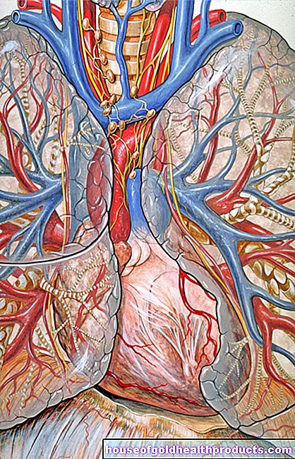
More about the experts All content is checked by medical journalists.The superior and inferior vena cava (superior vena cava, inferior vena cava) are the two largest veins in the human body. They collect oxygen-poor blood from the periphery of the body and direct it back to the heart, more precisely into the right atrium. Here you can find out everything you need to know about the paired vena cava!
What is the vena cava?
The superior and inferior vena cava (superior vena cava and inferior vena cava) are about two centimeters in diameter and are among the thickest veins in the body. Unlike the veins in the extremities, they have no venous valves that prevent the blood from flowing back. The two veins carry oxygen-poor, carbon-dioxide-rich blood from the periphery of the body back to the right heart, from where it is pumped further into the lungs to take in new oxygen and release carbon dioxide.
The name vena cava comes from the fact that the central veins in a corpse do not contain blood, so they are hollow inside (Latin: cava).
Superior vena cava
The superior vena cava (Vena cava superior) collects venous, deoxygenated blood from the veins of the head and neck area and the upper extremities and brings it back to the heart. It is about five to six inches long and runs along the right edge of the sternum.
Lower vena cava
The inferior vena cava (inferior vena cava) brings venous blood from the abdomen, pelvis, and legs to the heart. It runs to the right of the main artery (aorta) and runs through the diaphragm. It begins at the confluence of the right and left pelvic veins (which bring the venous blood out of the legs) and in the further course has inflows from the pelvic and abdominal organs: The venous blood from the unpaired abdominal organs (such as stomach, intestines, pancreas) is via the portal vein first transported to the liver before being fed into the inferior vena cava. The venous blood of the other organs in the abdomen and pelvis (such as the kidneys, testicles and ovaries) flows directly into the inferior vena cava.
Central venous catheter (CVC)
Due to their thickness and the fact that both vena cava flow into the right heart, a cardiac catheter (central venous catheter, CVC) can be pushed through a vena cava into the right atrium. This can be used to measure venous blood pressure within the vein, which doctors refer to as central venous pressure.It is normally at a maximum of 15 mmHg, but is subject to large fluctuations due to breathing and pulse conditions and can increase sharply in the event of a pulmonary embolism, for example.
A central venous catheter is not used to measure venous pressure - medication or infusions can also be administered via the thin plastic tube.
Diseases related to the vena cava
In pregnant women (especially when lying on their back), the heavy uterus can press on the inferior vena cava and thus restrict the return of blood to the heart. This vena cava compression syndrome (vena cava syndrome) triggers palpitations, drop in blood pressure, nausea and sweating and can also endanger the oxygen supply to the unborn child.
In addition, constrictions (stenoses), malformations, injuries as well as benign and malignant neoplasms of the vena cava can cause health problems.
Tags: therapies Diagnosis magazine