Humeral head fracture
Dr. med. Mira Seidel is a freelance writer for the medical team.
More about the experts All content is checked by medical journalists.A humeral head fracture (humerus head fracture, subcapital humerus fracture) is understood to be a fracture at the head of the humerus. This type of fracture is most common in the elderly with osteoporosis, usually caused by indirect trauma. A humeral head fracture is painful and restricts the mobility of the arm. Depending on the type of fracture, it is treated conservatively or surgically. Learn more about the humeral head fracture here.
ICD codes for this disease: ICD codes are internationally recognized codes for medical diagnoses. They can be found, for example, in doctor's letters or on certificates of incapacity for work. S42
Humeral head fracture: description
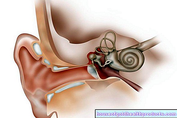
The humerus has a relatively large head that is three times larger than the socket in which it lies. This allows the shoulder a great deal of freedom of movement: the shoulder joint is the most flexible joint in the human body. The shoulder joint is mainly stabilized by the surrounding tendons, muscles, ligaments and soft tissues.
Structure of the humerus
The head (caput humeri) of the humerus is separated from the rest of the bone by a short ring-shaped neck (collum anatomicum). This is followed by two bony elevations that serve as attachment points for numerous muscles. The upper elevation is located on the outside of the humerus and is called the "greater tuberosity". The smaller elevation is called "Tuberculum minus".
A thinner neck (collum chirurgicum) follows directly below the lesser tuberosity. Here the bone is very soft and narrow. This point can break particularly easily when subjected to external force. The shaft of the humerus (humerus shaft) connects to the collum chirurgicum.
Fractures of the upper arm
The humeral head fracture is one of the proximal ("close to the body") humerus fractures. The upper arm can also break in other places. If this happens in the middle part of the bone, it is called a humeral shaft fracture. If, on the other hand, the bone breaks at its lower end, it is a distal humerus fracture.
Upper arm fractures near the shoulder joint make up about five percent of all fractures. This makes the upper arm the third most common fracture site in the human body. This fracture is common in old age. Women are two to three times more likely to be affected than men. In adolescents, such a break requires considerable force.
Humeral head fracture: classification
A humeral head fracture can break the head into various fragments. According to the doctor Codman, the humeral head fracture is divided into four main fragments with a typical direction of dislocation:
- Humeral head: tilting due to compression
- Greater tuberosity: the fragments are displaced backwards and upwards by pulling the muscles
- Lesser tuberosity: by pulling the muscles, the fragments are shifted towards the center front
- Shaft: by pulling the muscles, the fragments are shifted towards the center front
A classification of humeral head fractures according to the doctor Neer is based on the number of fragments with or without displacement:
- Group I: 1 fragment, no or minimal shift
- Group II: 2 fragments, displaced at the anatomical collar
- Group III: 2 fragments, tilted, displaced or the debris break
- Group IV: 2, 3 or 4 fragments, avulsion of the greater tuberosity, possibly avulsion of the lesser tuberosity
- Group V: 2, 3 or 4 fragments, avulsion of the lesser tuberosity, possibly avulsion of the greater tuberosity
- Group VI: dislocation fractures
A fragment is shifted more than a centimeter or twisted by more than 45 degrees.
An AO classification (Stans 2018) of proximal humerus fractures is based on the number of fragments:
- A: Extra-articular 2-fragment fracture
- B: Extra-articular 3-fragment fracture
- C: 4 fragment and articular fractures
Humeral head fracture: symptoms
If there is severe pain in the shoulder area after an accident, this may be an indication of a humeral head fracture. Another sign of such a break is an inability to move your arm or shoulder. The area is usually swollen and tender.
Furthermore, an extensive bruise (hematoma) forms as a result. After a day or two, it can sag to the elbow and cause the skin to discolour. In some cases, a malalignment of the upper arm is also visible in the case of an upper arm fracture.
Humeral head fracture: causes and risk factors
A humeral head fracture is usually caused by indirect trauma from a fall on the outstretched hand, elbow, or shoulder. In older people, osteoporosis (bone loss) plays an increasingly important role: Due to age-related hormone changes, the bone loses its strength, becomes porous and breaks more easily. Even harmless falls can then lead to a fracture, such as a fracture of the humeral head. About 70 percent of all patients with a humeral head fracture are older than 60 years.
A humeral head fracture is less common in young people than in older people and is often the result of serious traffic or sports accidents (rapid trauma). Babies can develop an upper arm fracture during childbirth.
Humeral head fracture: necrosis
The more severe the injury (especially in the area of the anatomical collum), the higher the risk of humeral head necrosis. In the process, the bone tissue on the head of the humerus dies. In the case of a humeral head fracture with additional dislocation (dislocation), the risk of necrosis is as high as 90 percent.
The reason for the humeral head necrosis is that the bone is no longer adequately supplied with blood. This happens when certain blood vessels are injured: the arteria circumflexa humeri anterior and its terminal branch the arteria arcuata and the arteria circumflexa humeri posterior. The humeral head necrosis is one of the aseptic, i.e. not infection-related bone necrosis.
Humeral head fracture: examinations and diagnosis
If a humeral head fracture is suspected, a doctor specializing in orthopedics and trauma surgery is the right contact. He will first ask you exactly how the accident happened and your medical history (anamnesis) and then examine you.
Medical history and physical examination
Possible questions from the doctor during the anamnesis interview are:
- Did you fall on your shoulder or outstretched arm?
- Can you describe the exact course of the accident?
- Can you still move your shoulder or arm?
- Do you have pain?
- Have you already had symptoms such as pain, restricted mobility or a previous dislocation in the shoulder or arm area?
A humeral head fracture can often be recognized from the description of the accident and the symptoms. Often the patient supports the injured arm on the wrist (in contrast to a broken shaft of the upper arm).
Similar symptoms to a humeral head fracture show a shoulder dislocation (shoulder dislocation). The doctor will therefore examine you for any nerve and vascular injuries that may be present.
Children who suffer from a birth-traumatic humeral head fracture often adopt a relieving posture. This is sometimes misinterpreted as plexus palsy (paralysis). This can be checked by means of a movement test: In the case of a broken upper arm, the child - in contrast to paralysis - has pain when the arm is moved.
Apparative investigations
To confirm the suspected diagnosis of a humeral head fracture, x-rays are usually taken of all sides of the shoulder. On the images, the doctor can also see whether parts of the fracture have shifted or whether other bony structures have broken.
If the fracture is only slightly displaced, the doctor checks whether the head fragments are stable even if the arm is carefully spread to the side by 80 degrees. Computed tomography (CT) provides more precise information, showing the exact relationship between the individual fragments. The CT examination is particularly indicated when an operation is to be planned.
For special questions, the doctor can order a magnetic resonance imaging (magnetic resonance imaging, MRI). In this way, for example, soft tissue damage such as tendon injuries can be detected or excluded.
Angiography (vascular x-ray) can be used to localize the location of a possible vascular injury. Electromyography (EMG) can be used to determine whether the muscles and / or nerves are still intact.
Humeral head fracture: treatment
There are different treatment measures depending on the severity of the humeral head fracture. In the case of an acute humeral head fracture, the most important thing is to treat the pain and avoid further damage. If a shoulder dislocation is suspected, no attempt should be made to straighten the joint again - this could cause even more damage! Imaging must always confirm the dislocation first.
Humeral Head Fracture: Conservative Therapy
In many cases, surgery can be avoided if the upper arm fractures are uncomplicated. If the fragments are not shifted against each other, the upper arm is usually immobilized with a special bandage (Desault or Gilchrist bandage) for about a week. Some patients receive accompanying cold therapy (cryotherapy).
The person concerned can then begin with light exercises, but these should not be done in the pain area. As soon as the pain subsides, physiotherapy begins with pendulum movements of the arm. After two to three weeks, the patient is allowed to move the arm actively and passively again.
It is important to observe the healing progress with X-ray controls. As a rule, a check-up follows after one day, ten days and six weeks. If the bone has healed adequately, it will be stable again after about six weeks.
Displaced fractures are only treated conservatively in rare cases. This is the case, for example, when there is a high risk of surgery. After applying special bandages, the arm is also immobilized with a plaster cast. A fracture of the upper arm can often be treated conservatively, especially in children, because it realigns itself spontaneously.
Humeral head fracture: operative therapy
In general, there are two different surgical procedures, depending on the location and the type of injury: osteosynthesis and joint replacement (endoprosthesis). The surgeon also decides, depending on the type of fracture, whether an open or a closed operation is indicated.
A humeral head fracture is basically an urgent but not an emergency operation. It should first be immobilized in the Gilchrist or Desault bandage and operated on within ten days.
If vessels or nerves have also been injured, an operation is usually performed immediately to avoid permanent damage.Even in the event of a dislocation that can no longer be straightened in, the doctor usually immediately decides to have an operation.
Osteosynthesis
In the case of a greater tuberosity fracture in which the fragments are displaced, the shoulder joint is often also dislocated. After the correction, the bone is then stabilized with plates, screws or drill wires. Then the arm is immobilized in a special bandage. If the fracture is not postponed, active muscle movement is recommended only after three weeks because of the high risk of displacement.
If it is an unstable humeral head fracture with a strongly displaced fracture or a dislocated fracture, surgery is also performed. The aim is to anatomically restore the humeral head in such a way that follow-up treatment is not necessary.
Endoprosthesis
Elderly people with poor bone quality and who are at high risk of bone necrosis are initially provided with an endoprosthesis. New angle-stable implants show good results. An endoprosthesis is also indicated, for example, if an osteosynthesis has failed, a dislocation fracture was a long time ago or the joint was necrotic destroyed.
In younger patients one always tries to preserve the humerus head and to realign the fracture parts anatomically.
Humeral head fracture: disease course and prognosis
It is recommended not to completely immobilize the shoulder joint for more than two to three weeks, as otherwise a so-called "frozen shoulder" can develop - a painful stiffening of the shoulder.
If a humeral head fracture is operated on, complications such as wound healing disorders, infections or secondary bleeding rarely occur. Occasionally a humeral head fracture does not heal completely (pseudoarthrosis). The function is hardly affected by this. The prognosis of a humeral head fracture is particularly good in children.
Other possible complications with a humeral head fracture are:
- Humeral head necrosis (especially in the elderly)
- Impingement: painful entrapment of soft tissues in the joint space (between the roof of the shoulder and the humeral head) in the case of a greater tubercle fracture
- Labral lesion (injury to the joint lip)
- Rotator cuff tear (tear in the muscle group in the shoulder area)
- Vascular and nerve damage (e.g. to the axillary nerves or the axillary artery) in severe humeral head fractures
The aim of the treatment is always that the upper arm is able to move in everyday life without pain. In some cases, however, the shoulder cannot move as it did before after a humeral head fracture. The arm can then not be moved forwards and sideways up to the vertical. This happens about 10 to 20 percent of the time.
Tags: first aid medicinal herbal home remedies stress


.jpg)