Hemolytic anemia
Astrid Leitner studied veterinary medicine in Vienna. After ten years in veterinary practice and the birth of her daughter, she switched - more by chance - to medical journalism. It quickly became clear that her interest in medical topics and her love of writing were the perfect combination for her. Astrid Leitner lives with daughter, dog and cat in Vienna and Upper Austria.
More about the experts All content is checked by medical journalists.Hemolytic anemia is when red blood cells are destroyed or broken down too soon. The causes are manifold. Typical symptoms are paleness, tiredness, yellowing of the skin and mucous membranes, and an enlarged spleen. Read here how hemolytic anemia works and what doctors do about it!
ICD codes for this disease: ICD codes are internationally recognized codes for medical diagnoses.They can be found, for example, in doctor's letters or on certificates of incapacity for work. D57D59D56D55D58

Brief overview:
- What is Hemolytic Anemia? Anemia due to the destruction or premature breakdown of red blood cells (erythrocytes)
- Course of the disease and prognosis: The course and prognosis depend on the underlying cause.
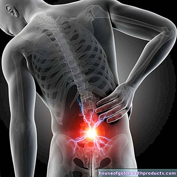
- Symptoms: paleness, weakness, circulatory problems up to fainting, headache, abdominal pain, back pain, yellowing of the skin and mucous membranes (jaundice), enlargement of the spleen (splenomegaly)
- Causes: congenital or acquired diseases, medication, drugs
- Diagnostics: Typical symptoms, blood test, blood smear, Coombs test, ultrasound, bone marrow biopsy
- Treatment: glucocorticoids (cortisone), immunosuppressants (drugs that reduce the immune system), bone marrow transplantation, removal of the spleen, administration of folic acid and iron
- Prevention: No specific preventive measures are possible.
What is Hemolytic Anemia?
Doctors call hemolytic anemia anemia, which is caused by the destruction or premature breakdown of red blood cells (erythrocytes). The breakdown and regeneration of red blood cells are normally subject to a cycle: healthy erythrocytes have a life cycle of around 120 days (70 to 90 days for newborns) before they are broken down and regenerated in the bone marrow.
In hemolytic anemia, this cycle is shortened: the red blood cells are broken down prematurely (on average after about 30 days), the new formation in the bone marrow lags behind. Overall, there are too few erythrocytes in the blood and breakdown products of the blood cells are deposited in the body. Typical signs of hemolytic anemia are paleness, tiredness, dizziness, circulatory problems, yellowing of the skin and enlarged spleen.
Haemolytic anemia has many possible causes, some are congenital (e.g. thalassemia, sickle cell anemia, spheroidal cell anemia, favism), others are only acquired in the course of life. Examples of acquired hemolytic anemias are autoimmune hemolytic anemia, rhesus intolerance, malaria, lead poisoning, incompatible blood transfusions, greatly increased copper levels and paroxysmal nocturnal hemoglobinuria.
Doctors also differentiate whether the cause of hemolysis lies in the red blood cells themselves (corpuscular anemia) or outside the blood cells (extracorpuscular anemia).
What is Hemolysis?
Hemolysis is the dissolution of red blood cells. This destroys the shell (cell membrane) that surrounds the red blood cells. Hemolysis in and of itself is not a disease; it takes place all the time in every body: red blood cells have a life cycle of around 120 days. Then they are dismantled and replaced with new ones. The bone marrow is responsible for the formation of new red blood cells: This is where new erythrocytes arise from precursor cells (megakaryocytes). The breakdown and regeneration of red blood cells usually go hand in hand. This ensures that sufficient blood cells are always available.
The regular breakdown of old erythrocytes takes place by so-called scavenger cells (macrophages) in the spleen, and to a lesser extent in the liver as well. They dissolve and break down the shell of the red blood cells. Macrophages occur in different tissues; doctors refer to them as a whole as the "reticuloendothelial system".
Erythrocytes have the task of binding oxygen to themselves and supplying it to the entire body. The main component of red blood cells is the red blood pigment hemoglobin. When the blood cells break down, the hemoglobin is broken down. The breakdown product bilirubin is produced, which reaches the bile via the liver and is excreted from there in the stool and, to a lesser extent, in the urine.
In hemolytic anemia, more red blood cells break down than usual, and at the same time the bone marrow lags behind in the formation of new cells. The result is that there are too few erythrocytes overall.
The liver is overwhelmed by the increased breakdown of red blood cells: so many breakdown products such as bilirubin accumulate that they can no longer be excreted quickly enough in the stool (and to a lesser extent in the urine). Instead, bilirubin builds up in the mucous membranes and skin. This leads to the typical yellowing of the skin and mucous membranes.
What is anemia
One speaks of anemia when the number of red blood cells (and thus also the red blood pigment hemoglobin) is below the age- and gender-specific reference value.
Anemia has many different causes: it is caused either by the rapid breakdown of red blood cells (hemolysis), enzyme or membrane defects in the blood cells, a distribution disorder (e.g. pregnancy anemia or hypersplenism due to an enlarged spleen) or an autoimmune reaction in which the immune system falsely Forms defense bodies (antibodies) against red blood cells. Other causes of anemia are certain chronic diseases or cancers.
Course of the disease and prognosis
Both the course and prognosis of hemolytic anemia depend on the underlying cause.
Autoimmune hemolytic anemia, for example, has a good prognosis, especially after the spleen has been removed. The same applies to congenital spheroidal cell anemia: after the spleen has been removed, the symptoms of the disease improve significantly. Overall, the treatment of congenital anemia has improved significantly over the past few decades. Secondary diseases and complications are largely manageable, and life expectancy has risen sharply. Older patients with additional cardiovascular diseases, in particular, have a less favorable prognosis.
In any case, it is important to have regular check-ups. In the case of chronic hemolysis, the doctor closely examines the blood.
What are the symptoms of hemolytic anemia?
There are different causes of hemolytic anemia. The symptoms that occur depend on the cause of the disease.
What they all have in common are the symptoms that are triggered by the excessive breakdown and breakdown of the erythrocytes. Its severity depends on how quickly the anemia develops: the faster, the more pronounced the symptoms. If the anemia is congenital or if it develops slowly, the body adapts to the anemia and sometimes develops no or only weak symptoms.
The following symptoms indicate hemolytic anemia:
- paleness
- fatigue
- Reduction in performance
- Low blood pressure (hypotension)
- dizziness
- headache
- tinnitus
- Circulatory problems up to fainting
- Palpitations
- Difficulty breathing
- Yellowing of the mucous membranes and skin (jaundice): caused by the breakdown of the red blood pigment (hemoglobin) contained in the red blood cells. The yellowish color is caused by bilirubin, the breakdown product of hemoglobin.
- Enlargement of the spleen (splenomegaly)
Possible complications of hemolytic anemia
Hemolytic crisis: A hemolytic crisis occurs when a large number of red blood cells dissolve in a short period of time. Such crises are possible with favism, sickle cell anemia and blood transfusions. Signs of a hemolytic crisis are:
- fever
- chills
- weakness
- Circulatory problems up to shock
- stomach pain
- Back pain
- headache
- Red or reddish-brown urine (when hemoglobin is excreted in the urine)
- Later yellowing of the mucous membranes and skin
A hemolytic crisis is a medical emergency. Call an emergency doctor at the first sign!
Gallstones: Gallstones develop in some patients as a result of chronic hemolytic anemia. They arise because bilirubin accumulates when the red blood pigment (hemoglobin) is broken down. It collects in the gallbladder and in some patients forms so-called "pigment stones".
Folic acid deficiency: the bone marrow needs folic acid in order to produce new, healthy red blood cells. A permanently increased formation of new erythrocytes can lead to a long-term folic acid deficiency.
Iron deficiency: If too many red blood cells are lost, iron deficiency occurs in the long term. Iron is part of the red blood pigment hemoglobin.
Cause and risk factors
Corpuscular hemolytic anemia
In corpuscular hemolytic anemia, the cause lies in the red blood cells themselves. This form of hemolytic anemia is usually inherited and manifests itself in childhood or adolescence. Different blood cell defects trigger hemolytic anemia here:
- Congenital cell membrane disorder: spheroidal cell anemia (hereditary spherocytosis)
- Acquired cell membrane disorder: paroxysmal nocturnal hemoglobinuria
- Disorder of the erythrocyte metabolism: favism (glucose-6-phosphate dehydrogenase deficiency)
- Hemoglobinopathy: sickle cell disease, thalassemia
Extracorpuscular hemolytic anemia
Extracorpuscular anemia is not caused by the red blood cells themselves, but by external factors. This form of hemolytic anemia is usually not congenital, but rather develops over the course of life.
Possible causes are:
- Medicines: Some active ingredients such as quinine and mefloquine (antimalarials), penicillin, antibiotics such as metronidazole, psychotropic drugs such as bupropion or pain relievers (NSAIDs) such as ibuprofen can trigger hemolytic anemia.
- Autoimmune diseases: In the case of an autoimmune disease, the immune system falsely forms defenses (antibodies) against harmless endogenous substances, for example against red blood cells. Examples are immune thrombocytopenia (ITP) and autoimmune hemolytic anemia of the cold type.
- Infections: In some cases, infections cause hemolytic anemia. The most common pathogens include Clostridium perfringens, streptococci, meningococci, plasmodia, Bartonella, Epstein-Barr virus and mycoplasma.
- Mechanical injuries to the erythrocytes: Here the red blood cells are damaged and destroyed by mechanical obstacles in the blood circulation (e.g. artificial heart valves).
- Enlargement and increased activity of the spleen (hypersplenism): When the spleen enlarges, more blood cells are broken down.
- Poisons (toxins): Poisoning with lead or copper leads to the fact that more red blood cells are broken down.
- Drugs: Drugs like ecstasy or cocaine can cause hemolytic anemia.
Investigation and diagnosis
Congenital hemolytic anemias such as sickle cell anemia or globular cell anemia are usually diagnosed in childhood or adolescence. In many cases, doctors only discover acquired anemia by chance during blood tests, for example during a preventive medical check-up. If the haemolytic anemia is already advanced, the yellowing of the skin or blood in the urine is usually the reason for the first visit to the doctor. The first point of contact is, depending on the age of the patient, the pediatrician or the family doctor.
anamnese
In the initial consultation, the doctor asks about the current symptoms and asks how long they have been present. If haemolytic anemia is suspected based on a blood test, the doctor will inquire about further abnormalities. These include:
- Family History: Has there been a family history of hemolytic anemia (such as thalassemia, sickle cell anemia, or favism)?
- Do you have a fever or other symptoms?
- Is the patient taking any medication? If yes, which?
- Does the patient take drugs? If so, which ones (especially cocaine)?
Blood test
If no current blood results are available, the doctor will take blood from the patient and pay particular attention to the following values:
- Low numbers of red blood cells (erythrocytes) and red blood pigment (hemoglobin)
- Increased number of reticulocytes (reticulocytosis, precursor cells of the red blood cells in the bone marrow)
- Low haptoglobin (transport protein for the red blood pigment hemoglobin)
- Increased bilirubin (the pigment of bile, a sign of increased hemoglobin breakdown)
- Elevated LDH (signs of increased cell breakdown)
- Folic acid or iron deficiency
Blood smear
For a blood smear, the doctor puts a drop of blood on a glass slide and examines the individual blood cells under the microscope for changes. Certain changes in the shape of the red blood cells indicate specific diseases that cause hemolytic anemia. In spherical cell anemia, for example, the erythrocytes are spherical instead of flat.
Urinalysis
The main part of the red blood pigment - or its breakdown product bilirubin - is excreted in the stool, a small part in the urine. If bilirubin is found in the urine, it is a sign of an increased breakdown of red blood cells. Doctors speak of "urobilinogen".
Coombs test
The Coombs test is a blood test that detects antibodies against red blood cells. The doctor uses this to check whether there are antibodies against the red blood cells in the patient's blood.
Ultrasound examination
An ultrasound scan of the abdomen will reveal whether the spleen and / or liver are enlarged.
treatment
How the doctor treats hemolytic anemia depends on the underlying cause. Regardless of the cause of the disease, the goal of treatment is to eradicate the anemia and control the disease.
Glucocorticoids and immunosuppressants: Glucocorticoids (cortisone) and immunosuppressants (drugs that suppress an excessive reaction of the body's own immune system) help with autoimmune hemolytic anemia.
Removal of the spleen (splenectomy): When the spleen is removed, fewer red blood cells are broken down. This helps alleviate the symptoms, but makes people more susceptible to infections. For this reason, patients are vaccinated against certain diseases (such as influenza, pneumococcal, meningococcal and Haemophilus influenzae infections) a few weeks before the operation.
Avoidance of triggering drugs: If the cause of the hemolytic anemia lies in a certain active ingredient, the doctor will change the drug and, if necessary, switch to another, equivalent preparation.
Folic acid: In hemolytic anemia, the body needs more folic acid to stimulate the production of red blood cells in the bone marrow. Doctors recommend a diet high in folic acid including spinach, lettuce, cabbage, fruit, whole grains, wheat germ, soybeans, legumes, and liver. If this is not enough, it makes sense to take a folic acid supplement.
Bone marrow transplant: Sickle cell anemia and thalassemia can be cured with a bone marrow transplant. Bone marrow is transferred from a healthy donor to the patient.
Phototherapy: Infants born with favism or rhesus intolerance may benefit from phototherapy. The babies are irradiated with short-wave light, which converts the bilirubin in the skin and then excretes it through the bile and kidneys.
Protection from the cold: In chronic autoimmune hemolytic anemia of the cold type, the most important measure is to protect those affected from the cold.
Prevent
Since hemolytic anemia has various causes, it can only be prevented to a limited extent. In any case, it is important to counteract nutritional deficiency symptoms. Eat a balanced diet and consume enough folic acid - contained in spinach, beans, cabbage and liver. The same applies to vitamin B12: it is contained in fish, dairy products, meat and eggs. Doctors recommend that women consume foods containing iron during menstruation. Red meat, whole grain products, legumes and nuts are ideal.
Tags: alcohol eyes laboratory values