Esophageal cancer
Ricarda Schwarz studied medicine in Würzburg, where she also completed her doctorate. After a wide range of tasks in practical medical training (PJ) in Flensburg, Hamburg and New Zealand, she is now working in neuroradiology and radiology at the Tübingen University Hospital.
More about the experts All content is checked by medical journalists.Cancer of the esophagus (esophageal carcinoma) is a particularly insidious cancer: since the cancer only causes symptoms such as difficulty swallowing at an advanced stage, it is usually discovered late. As with almost every cancer, a late diagnosis worsens the chances of survival - in the case of esophageal cancer even considerably. The two most common forms of esophageal cancer are squamous cell carcinoma and adenocarcinoma, which develop from different cell types. Here you can read everything you need to know about esophageal cancer.
ICD codes for this disease: ICD codes are internationally recognized codes for medical diagnoses. They can be found, for example, in doctor's letters or on certificates of incapacity for work. C15

Esophageal cancer: description
Esophageal cancer is a relatively common cancer worldwide. However, esophageal cancer is rather rare in Germany. According to the Center for Cancer Registry Data at the Robert Koch Institute in Germany, around 1,000 women and 4,000 men develop it every year. The mean age of onset is 66 years. Esophageal cancer before the age of 40 is rare. The number of new cases each year (incidence) has been increasing continuously since the 1980s. In women in particular, the number of new esophageal cancer cases is rising significantly.
Doctors assume that the steadily increasing cases of esophageal cancer in the last few decades can be attributed to lifestyle factors such as overeating and the consumption of alcohol and nicotine. These factors favor the so-called reflux disease. Reflux means that acidic gastric juice gets into the esophagus and damages the mucous membrane there. Reflux disease is significantly involved in the development of adenocarcinoma of the esophagus. Adenocarcinoma is currently only the second most common form of esophageal cancer, but overall this form is occurring more and more often and is a major contributor to the overall increasing number of esophageal cancer.
Life expectancy and chances of recovery depend on how far the cancer is when it is discovered. Most of the time, esophageal cancer is unfortunately only diagnosed late, when it has already spread to the surrounding lymph nodes and neighboring organs (metastasis). At the time of diagnosis, only about 40 percent of those affected can be helped with an operation. Although the prognosis of esophageal cancer has improved significantly in the last few decades thanks to today's therapeutic options, many people die from the tumors. Of the patients diagnosed with esophageal cancer, only about 15 to 20 percent survive the next five years.
Esophageal cancer can develop anywhere in the esophagus. However, cancer is more common in three areas of the esophagus. These are sections in which other organ structures constrict the esophagus somewhat: the entrance of the esophagus directly behind the pharynx, in the area where the esophagus passes the aortic arch and when the esophagus passes through the diaphragm. Depending on the type of degenerated cell, esophageal cancer is divided into different histological forms:
Esophageal cancer: squamous cell carcinoma (about 80 percent)
In squamous cell carcinoma, tumor cells develop from cells in the mucous membrane (squamous epithelium) of the esophagus. This type of cancer can develop in any part of the esophagus. About 15 percent occur in the first third of the esophagus, 50 percent in the middle and 35 percent in the last third. Squamous cell carcinoma is favored by heavy alcohol consumption, hot drinks, smoking and fungal toxins.
Esophageal cancer: adenocarcinoma (about 20 percent)
In an adenocarcinoma, the tumor arises from altered gland cells. It forms in the lower part of the esophagus in 95 percent of cases. The main cause of this is reflux disease, in which acidic stomach contents repeatedly enter the esophagus. It damages the mucous membrane, which initially leads to cell changes, a so-called Barrett's esophagus, which eventually develops into Barrett's carcinoma (adenocarcinoma). Adenocarcinomas have increased exponentially over the past few decades.
Esophageal cancer: undifferentiated carcinoma (about 10 percent)
If the original cell type from which the tumor developed can no longer be determined with certainty, doctors refer to this as "undifferentiated esophageal carcinoma". It is the rarest form of esophageal cancer.
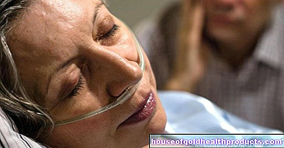
Esophageal cancer: symptoms
You can read everything you need to know about the typical signs of esophageal cancer in the article Esophageal cancer - symptoms.
Esophageal cancer: causes and risk factors
There are different risk factors for the two main forms of esophageal cancer (squamous cell carcinoma and adenocarcinoma):
Squamous cell carcinoma risk factors: Esophageal cancer, which develops from degenerate cells of the squamous epithelium, is primarily caused by
- Consumption of high proof alcohol
- smoking
- Consumption of hot drinks
- Nitrosamines (found in many foods)
- Aflatoxins (poison from mold)
- Achalasia (when the lower esophageal sphincter does not relax enough to allow food to pass easily)
Adenocarcinoma risk factors: Five percent of people with reflux disease (chronic heartburn) develop what is known as Barrett's esophagus, which is a precancerous stage. The normal mucous membrane cells of the esophagus are transformed into gland cells (metaplasia). Ten percent of this preliminary stage results in an adenocarcinoma of the esophagus, which is then also referred to as Barrett's carcinoma.
There are other factors that contribute to all forms of esophageal cancer. These include:
- Previous radiation therapy near the esophagus (for example, for breast cancer)
- HPV 16 papillomavirus infection (also involved in cervical cancer)
- Genetically caused corneal thickening of the hands and feet (tylosis palmaris and plantaris)
- Narrowing scarring after caustic burns
- Plummer-Vinson syndrome: a rare condition caused by iron deficiency
In addition to the listed possible causes of esophageal cancer, there are also protective factors. Studies have shown that people who take acetylsalicylic acid (Aspirin®, “ASA”) or other active ingredients from the group of non-steroidal painkillers for a long time seem to develop esophageal cancer less often. However, you should not take such drugs preventively, as they can cause serious side effects such as stomach ulcers on their own.
Esophageal cancer: examinations and diagnosis
The right contact for suspected esophageal cancer is a specialist in internal medicine who specializes in diseases of the digestive tract (gastroenterologist). The doctor first inquires about your current symptoms and any previous illnesses (anamnesis). Basically, the symptoms of esophageal cancer only appear at a very advanced stage of the disease ("silent cancer"). For example, your doctor may ask you the following questions if you suspect esophageal cancer:
- Have you accidentally lost weight in the past few weeks or months?
- Do you suffer from loss of appetite and nausea?
- Do you have pain when swallowing or a feeling of pressure in the throat or behind the breastbone?
- Did you vomit?
- Do you take any medicine?
Even if you have already developed esophageal cancer, such symptoms often only appear sporadically or not at all. Your doctor will also try to clarify the risk factors for esophageal cancer mentioned above. If, during an examination or in your medical history, there are suspicions of esophageal cancer, your doctor will refer you to a gastroenterologist.
The anamnesis is followed by a physical examination. The doctor checks whether the lymph nodes are enlarged or whether there are nodes elsewhere. Since the esophagus can only be assessed to a very limited extent from the outside, further examinations are usually necessary if esophageal cancer is suspected.
Further investigations
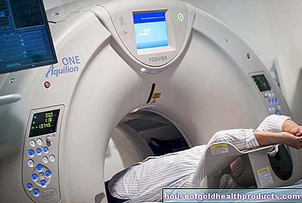
The esophagus and the ability to swallow can be assessed on the basis of various tests. This includes esophagoscopy, an ultrasound of the esophagus (endosonography) and the so-called X-ray swallow. In the latter case, the patient ingests a contrast medium that enables the doctor to follow the swallowing process precisely. Other imaging methods such as computed tomography (CT), magnetic resonance tomography (MRT) or positron emission tomography (PET) can be used to determine the spread of the tumor in the body (staging). Depending on the result of the staging, a stage-dependent therapy is carried out.
Esophageal cancer: esophagoscopy
An esophagoscopy is an examination of the esophagus. Similar to a gastroscopy, the person examined must be sober. Before the examination, he is given a light sleeping pill so that he does not consciously experience the examination himself and does not feel any pain. For the examination, a tube with a small camera and a light is passed through the mouth and into the esophagus. The doctor can see on a screen whether the lining of the esophagus has changed or whether it appears narrowed in certain places. If a certain area looks different, he can take a tissue sample (biopsy) with small forceps. This usually takes place in several places. The samples obtained in this way are then examined under the microscope. With the histological examination of the tissue samples, cancer precursors such as Barrett's esophagus can also be determined.
Esophageal cancer: endoscopic ultrasound
Endoscopic ultrasound of the esophagus is similar to esophagoscopy in its implementation. In this case, however, an ultrasound head is inserted into the esophagus. With the help of this method, the extent of the affected areas can be estimated well - an important piece of information in the case of esophageal cancer. The prognosis and therapy depend largely on whether the esophageal cancer is already affecting deep tissue layers and whether it has already spread to surrounding structures (e.g. lymph nodes). In addition, enlarged lymph nodes can be detected by endoscopic ultrasound.
Esophageal cancer: X-ray swallow
In the so-called X-ray swallow, the patient is asked to swallow an X-ray contrast medium. X-rays are taken while swallowing. Instead of a single X-ray, this examination provides a short film in which the swallowing movement and also the size and shape of the esophagus can be assessed. With the help of the X-ray swallow, for example, constrictions (stenoses), asymmetries or changes in the contour of the esophagus can be detected. Such changes can indicate esophageal cancer.
Esophageal cancer: imaging
In order to determine the exact spread of esophageal cancer in the body (staging), computer, magnetic resonance or positron emission tomography is usually carried out. For these examinations, the patient is driven on a couch into a tube in which images of the affected parts of the body are created. Each of these studies has advantages and disadvantages. That is why the doctor decides individually which of the procedures makes the most sense. Sometimes the patient is given a contrast medium to drink before this examination.
The aim of these various procedures is to determine exactly where the tumor is located, which neighboring structures it is adjacent to and how large it is. The decisive factor is whether the esophageal cancer has already spread to lymph nodes or other organs. This information determines the subsequent therapy for esophageal cancer. The smaller the tumor, the greater the chances of recovery - and the less it has already spread. Even in the case of an advanced disease, a detailed examination can provide more targeted treatment and improve the esophageal cancer prognosis.
Esophageal cancer: complementary diagnostics
If it is suspected that the esophageal cancer has already grown into the larynx or the bronchi, a mirroring of these organs is necessary (bronchoscopy, laryngoscopy). The examination is similar to a gastroscopy. However, a slightly thinner hose is used for this. In other cases, an ultrasound of the upper abdomen is performed.
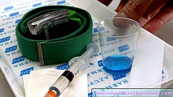
If the CT, MRI, PET or X-ray images show abnormal structures in the bone, a bone scintigraphy is useful. For this purpose, a contrast agent is injected into the patient's arm vein, which is mainly deposited in the bone in particularly metabolically active, well-perfused areas. This also applies to metastases. Sub-tumors (metastases) then appear as dark spots on images taken by a so-called gamma camera.
Esophageal cancer: treatment
Various methods are available for treating esophageal cancer. Surgery, radiation or chemotherapy - which therapy option is used depends on the size of the tumor, whether it has spread in the body and the general condition of the patient. Often, different procedures are combined. A special case are patients whose esophagus is so narrowed by the tumor that they can no longer take in food. In this case, the doctor can stretch the esophagus (bougienage) and insert a metal tube (stent) that keeps the path open for food.
Esophageal cancer: surgery
If it is discovered at a very early stage, the chances of a cure for esophageal cancer are very high. In many cases, the tumor can then simply be removed as part of an esophagoscopy (endoscopic). If the tumor is a little more advanced, a major intervention is necessary. The entire esophagus is removed along with the associated lymph nodes. So that patients can eat again after the operation, the doctor inserts a piece of the small intestine in place of the esophagus. Alternatively, he can sew the stomach directly to the upper part of the esophagus (stomach pull-up). If the surgeon succeeds in removing the tumor completely and he has not yet spread any metastases, this intervention can be sufficient for complete healing up to stage IIa.
Esophageal cancer: chemotherapy and radiation
In advanced esophageal cancer, chemotherapy or radiation therapy in addition to surgery has proven useful. In some cases, chemotherapy or radiation is carried out before the operation to reduce the size of the tumor ("downstaging"). As a result, the surgical procedure should be less serious, which reduces the surgical risk for the patient. Despite this combined therapy, only 35 percent of those affected survive a completely removed esophageal carcinoma. Scientific studies are currently testing whether chemotherapy should be combined with radiation to improve the prognosis for esophageal cancer. This form of therapy (chemotherapy + radiation) is also chosen if an operation cannot be performed.
Esophageal cancer: palliative therapy
An esophageal cancer that is very advanced can no longer be cured. So that the patient has as few complaints as possible, palliative (symptom-relieving, non-healing) therapy is carried out. It is determined individually and can include chemotherapy or radiation. In end-stage esophageal cancer, the patient can also receive extra food through a gastric tube.
Esophageal cancer: disease course and prognosis
Esophageal cancer grows quickly into the surrounding organ structures. First, it expands into the outer layers of the wall of the esophagus. In the end-stage esophageal cancer, the lung mantle, the heart mantle, the diaphragm, the main artery (aorta), the vertebral bodies or the trachea can be affected. Before the tumor affects these other organs, however, it usually first spreads into the lymph nodes. Cancer cells spread via the blood vessels and also settle in the liver, lungs, brain or bones. Such metastases are unfortunately not uncommon in esophageal cancer.
Life expectancy and prognosis for this tumor disease are unfortunately still poor overall. This is mainly due to the fact that esophageal cancer is usually only discovered when the tumor is already relatively large and has spread to the lymph nodes. This is the case for 90 percent of patients. The more advanced the disease, the worse the prognosis. At a very early stage, the tumor can be removed and healed using an esophagoscopy or surgery.
If the cancer has already spread, only 35 percent of patients will survive the next five years - even if the tumor has been completely surgically removed and then received chemotherapy or radiation. If the operation is dispensed with and only chemotherapy and radiation are carried out, 30 percent will survive the next three years. Patients at increased risk should therefore be examined regularly by an internist so that he can detect esophageal cancer in good time.
Tags: foot care therapies organ systems


.jpg)


.jpg)