Hematoma (bruise)
Martina Feichter studied biology with an elective subject pharmacy in Innsbruck and also immersed herself in the world of medicinal plants. From there it was not far to other medical topics that still captivate her to this day. She trained as a journalist at the Axel Springer Academy in Hamburg and has been working for since 2007 - first as an editor and since 2012 as a freelance writer.
More about the experts All content is checked by medical journalists.
Whether in sport or everyday life - a hematoma (bruise) is easy to deal with. If you bump against the edge of the table or fall, a blood vessel can tear and bleed into the surrounding tissue - the typical "bruise" forms. Read here what other forms of hematoma there are, when a disease can be the cause and how a bruise can be treated!

Brief overview
- What is a hematoma? Bleeding into tissue or a body cavity as a result of blood leaking from injured vessels.
- Forms of hematoma: e.g. subcutaneous hematoma (hemorrhage under the skin, the typical "bruise"), intramuscular hematoma (hemorrhage in muscles), intracranial hematoma (bruise in the skull), subungual hematoma (bruise under a nail), hemarthrosis (bruise in one joint), eyeglass hematoma (bruise around both eyes)
- Development: Hematomas usually arise as a result of trauma such as a fall or impact injury, sometimes also after surgery, in certain diseases (such as hemophilia, myelodysplastic syndrome) or treatment with anticoagulant drugs. Spontaneous hematomas are also possible.
- Symptoms: typical color gradient in superficial hematomas (from red to yellow-brown), pressure pain, depending on the location further / other symptoms such as swelling, impaired joint function (e.g. with hemarthrosis), headache and disturbed attention (with bruise in the skull) etc.
- Treatment: Not necessary for normal "bruises", if necessary cool, store high, heparin or arnica ointment. Larger hematomas and those that cause complications (e.g. pressure on nerves) are surgically removed.
What is a hematoma?
A hematoma is a collection of blood in tissue or in a preformed body cavity, such as a joint space. The blood comes from injured blood vessels. Colloquially, a hematoma is also called a "bruise".
Forms of hematoma
The hemorrhage can occur in very different body tissues and regions, which is reflected in the medical name. Here are some examples:
Subcutaneous hematoma
The typical "bruises" are bleeding under the skin (in the subcutis) and are called subcutaneous hematomas in technical terms. They are usually the result of blunt trauma, such as a shock injury. But even after a blood sample has been taken, a "bruise" can develop at the puncture site.
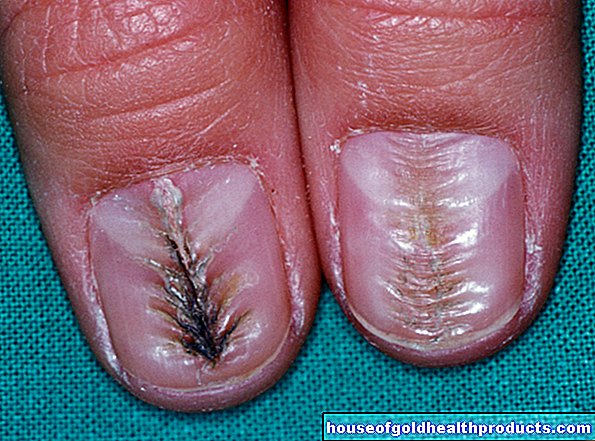
Subungual hematoma
If you pinch your hand by a door or a drawer, a subcutaneous bruise can develop on the finger or fingers. However, if a fingernail is caught while being trapped and bleeds under it, there is a subungual hematoma ("ungus" = Latin for nail). This can also occur if you hit your fingernail with a hammer or repeatedly wear shoes that are too tight.
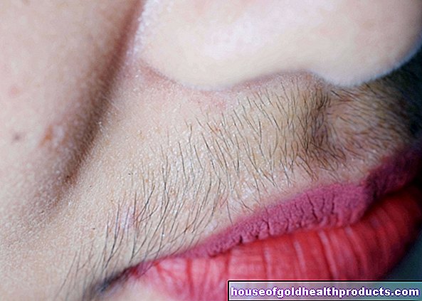
Bruise on the face
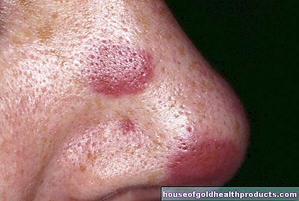
A bruise on the face often looks dramatic. If it develops in a ring around an eye (e.g. as a result of a punch), doctors speak of a monocular hematoma. If there is a ring-shaped bruise around both eyes, the diagnosis is ophthalmic hematoma. Both forms of hematoma can be signs of a skull fracture!
Sometimes there is also a bruise in the eye itself, for example through bleeding under the conjunctiva - called hyposphagma. It occurs when a small vein of the conjunctiva bursts, which can happen, for example, with severe coughing or straining (such as during childbirth), rapid changes in external pressure (when diving or on a plane) or high blood pressure.
Hematoma in the periosteum
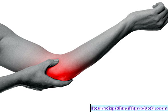
If you get a bump or blow to the shin, it usually triggers a hematoma in the periosteum. The periosteum is the thin layer of tissue with which all bones are covered on their outside (except in the area of the cartilaginous joint surfaces). Bleeding into this tissue occurs preferentially on the shinbone, because this bone is only covered by a thin layer of skin and hardly any fat or muscle tissue.
Hematomas in muscles
If the blood from injured blood vessels spills into muscle tissue, an intramuscular hematoma results. Bleeding into a muscle box can have serious consequences. This is a group of muscles that is enclosed by a thin, barely stretchable "skin" (fascia). The leg and arm muscles in particular are made up of such delimited compartments. If blood from injured vessels collects in a box, it can exert dangerous pressure on the muscle tissue and the (intact) vessels and nerves running here (compartment syndrome). In the worst case, the muscle tissue can die as a result.
Hemarthrosis
Doctors refer to hemarthrosis as bleeding into a joint, i.e. a bloody joint effusion. For example, if you fall badly (for example while skiing) you can get a bruise in your knee.
Hematoma in the skull
Cerebral hemorrhage can cause dangerous bruising within the skull - intracranial bruises. For example, blood can accumulate between the skull bones and the hard meninges (dura mater) to form what is known as an epidural hematoma (epidural hematoma). If there is bleeding into the subdural space (= gap-shaped space between the hard meninges and the middle meninges), the doctor diagnoses a subdural hematoma (subdural hematoma). A bruise right in the brain is called an intracerebral hematoma.
Hematomas in the skull can put dangerous pressure on the sensitive brain tissue. If there is no rapid relief, nerve cells can die.
How does a hematoma develop?
Most hematomas result from trauma. In the event of a fall, impact or blow, for example, blood vessels can tear and bleed into the surrounding tissue or into neighboring body cavities (such as a joint). Sometimes these bruises accompany other injuries such as bruises, bruises, or broken bones.
You can also develop bruises after surgery, for example if vessels were injured during the procedure.
Sometimes a hematoma develops spontaneously without a trigger. For example, the above-mentioned hyposphagma - a bruise in the eye under the conjunctiva - can occasionally occur for no particular reason. It is then referred to as "idiopathic".
Disease-related tendency to hematomas
Some people get noticeably easy bleeding (such as nosebleeds) and seemingly "bruises" for no reason. Causes can be diseases, for example a (as yet undiscovered) blood clotting disorder. This can have a wide variety of triggers, for example hemophilia or severe liver disease.
Blood clotting can also be specifically disturbed by medication: Anticoagulants such as phenprocoumon or heparin are used to prevent diseases that are caused by blood clots (e.g. stroke, heart attack). A side effect of treatment is that people may easily develop bleeding and bruising.
The so-called myelodysplastic syndrome (MDS) can also be responsible if someone is prone to bleeding and "bruising" for no reason or if they suffer from minor trauma (e.g. a small bump). MDS comprises a group of diseases in which the formation of various blood cells is impaired - including those of the blood platelets (thrombocytes). These are important for blood clotting.
Hematoma: symptoms
A simple, superficial hematoma is visible as a more or less tender "bruise" on the skin. Depending on the depth of the bleeding, the discoloration of the skin can become visible very quickly or only after hours or days. Blue is not the only color that a hematoma takes on: a superficial bruise changes its color over time from red to yellow-brown (see below).
A deep hematoma is usually not noticeable as a skin discoloration, but rather as a swelling. The congested blood puts pressure on the surrounding tissue, which is often painful. Extensive collections of blood can also affect the functioning of muscles and joints.
In the worst case, a hematoma located deep in the muscle tissue cannot be completely dissolved (resorbed). Over time it becomes encapsulated and calcified. Such an encapsulated, hardened hematoma can be very painful and also impede the function of muscles and joints.
Some hematomas can also cause specific symptoms depending on their location. For example, bleeding under the conjunctiva (hyposphagma) shows up as a sharply defined red spot in the white of the eye. It is usually harmless - does not hurt and does not impair vision. A bruise under a nail (subungual hematoma) manifests itself as a purple to black spot under the nail, which can be associated with severe throbbing pain.
A hematoma in the skull (intracranial hematoma) must be taken seriously. Among other things, it can make itself felt with headaches and impaired alertness and ability to react (vigilance disorder), for example because the leaked blood presses on brain tissue - nerve cells can die!
Very extensive hematomas can also be dangerous - the large amount of blood loss can trigger symptoms of shock such as restlessness, pale skin, tremors and cold sweat. This often happens with injuries to the thigh (large leg artery!).
If there are signs of a hematoma in the skull or a shock, you must call the emergency doctor quickly! There is danger to life!
Hematoma: course
The visible color change of superficial hematomas shows the healing process, i.e. the breakdown of the blood that has passed into the tissue:
- Red signals the bleeding into the tissue - the red blood pigment hemoglobin is responsible for the red color.
- Dark red-blue indicates the onset of blood clotting.
- Brown-black indicates that proteins have broken down the blood pigment into the pigment verdoglobin (choleglobin).
- A bruise glows dark green as soon as the verdoglobin has broken down into biliverdin.
- Yellow-brown is the final stage of a hematoma. The color is caused by the conversion of biliverdin to bilirubin - the end product of the breakdown of hemoglobin, which the body can transport to the liver via the bloodstream. Together with the bile produced there, the bilirubin can be excreted through the intestine.
How long does a "bruise" take to heal?
The healing process of a normal hematoma - i.e. the breakdown of the leaked blood and the resorption of the end products - usually takes two to three weeks. With larger hematomas, it can take longer, which is why you sometimes "help" by removing the bruise (see below: treatment).
If there is a large bruise under a fingernail or toenail, it usually falls off after a few weeks because the blood separates the nail from the nail bed. If, on the other hand, the subungual hematoma is only small, the nail usually stays on. However, it can take several weeks for the blackish discolored nail to grow out.
Hematoma: diagnosis
"Bruises" on the leg, arm, head or any other visible part of the body can be recognized as hematomas at first glance. Further medical examinations are generally not necessary unless the hematoma is very extensive or the doctor suspects additional injuries.For example, if the bruise could be connected to a broken bone, an X-ray examination will provide certainty. An ultrasound can make a bruise in the knee or in deeper tissue layers visible.
A more detailed diagnosis is also particularly necessary in the case of a bruise in critical areas (such as in the eye area) and if internal bleeding and hematomas are suspected (e.g. in the skull). For example, a hematoma in the skull can be detected using computed tomography (CT).
A doctor will also investigate more closely if someone often gets bruises for no reason and with minor injuries and is prone to bleeding (such as nosebleeds) and prolonged bleeding times. He will then ask if the patient is taking any medications - they may be drugs that inhibit blood clotting. Blood tests can also be instructive. For example, the number of blood platelets (thrombocytes) and blood clotting parameters (such as the Quick value or INR) may indicate a blood clotting disorder.
Hematoma: treatment
Simple "bruises" are harmless and will go away on their own after a few days. The following first aid measures are recommended if, for example, a ball hits your upper arm on the tennis court, your lower leg bumped against the table leg or you fell roughly on your bottom while ice skating:
- Cool the injured area for about 15 to 20 minutes, for example with ice or a cool pack (do not put either directly on the skin, but rather wrapped in fabric). You can also make an envelope with cold water. The cold causes the blood vessels to contract, which reduces the bleeding into the tissue. In addition, cold has a pain-relieving effect.
- If the swelling is large, you should elevate the injured body region if possible and keep it still. This also reduces blood flow to the injured area.
- A heparin ointment helps against bruises and swelling. You can also treat a bruise with a pain-relieving arnica ointment.
In general, the PECH scheme is recommended for first aid for sports injuries: break, ice, compression (pressure bandage), elevated position. Read more about it here.
Very large and rapidly spreading hematomas need medical attention as soon as possible. The loss of blood can cause a life-threatening shock! In addition, a large accumulation of blood can lead to infections and, depending on the location, press on sensitive tissue - for example an intracranial hematoma on the brain tissue, a bruise in the eye socket on the eyeball, optic nerve and / or eye muscles. In such cases, surgery is often used. The doctor removes the clotted blood in order to reduce the pressure on the surrounding tissue.
If a hematoma has hardened, hurts and maybe even affects muscles or joints in their functionality, it is also removed in one operation.
A bloody joint effusion (hemarthrosis) is also cleared out. The still liquid blood in the knee, for example, can be drained by means of a joint puncture. To do this, the doctor places a thin plastic tube through which the blood can drain (drainage).
If there is a bruise under a nail (subungual hematoma), the doctor may burn a small hole in the nail plate to drain the blood. For this he usually uses a needle or a heated wire, so it's quick and hardly hurts.
Medical help is always advisable in the following cases: large bruises, severe impact or blow injuries to the head or genitals, bruises in the eye area, severe swelling or pain, restricted mobility of muscles or joints, suspected further injuries (such as broken bones, joint damage), signs for a shock.
Treat the cause of the disease
If an illness is the reason if someone repeatedly gets "bruises" on the legs, arms or other parts of the body, this will be treated specifically depending on the possibility and need. For example, many hemophilia patients are given factor concentrates - concentrates of coagulation factors - via the vein (intravenously) to reduce the tendency to bleed. If a coagulation disorder is based on a vitamin K deficiency (for example in chronic liver diseases), this can be compensated for with medication.
Hematoma: prevention
You often get "bruises" while exercising. With protective equipment, you can prevent bruises and other injuries. For example, a protective helmet is advisable when cycling, motorcycling and on the ski slopes. Protective goggles protect sensitive eyes from injuries. Knee and elbow protectors are especially important for inline skaters, with shin guards passionate football players are well advised.
If you use the above mentioned first aid rules immediately after a shock, blow or fall injury (cool, store up, pause, use heparin or arnica ointment, etc.), you may be able to avoid developing a hematoma or this at least keep it small.
Are you prone to bleeding and bruising due to an illness or treatment with anticoagulants? Then you should refrain from injurious sports and be particularly careful in everyday life, for example on icy roads in winter.
Tags: sports fitness palliative medicine diet