Uveitis
and Sabine Schrör, medical journalistRicarda Schwarz studied medicine in Würzburg, where she also completed her doctorate. After a wide range of tasks in practical medical training (PJ) in Flensburg, Hamburg and New Zealand, she is now working in neuroradiology and radiology at the Tübingen University Hospital.
More about the expertsSabine Schrör is a freelance writer for the medical team. She studied business administration and public relations in Cologne. As a freelance editor, she has been at home in a wide variety of industries for more than 15 years. Health is one of her favorite subjects.
More about the experts All content is checked by medical journalists.Uveitis is inflammation of the middle skin of the eye (uvea). This consists of different sections that can be affected individually or in combination. Accordingly, there are different forms of uveitis. The disease can be caused by pathogens or occur in connection with another disease. However, the cause often remains unknown. Read everything you need to know about the causes, symptoms, and treatment of uveitis.
ICD codes for this disease: ICD codes are internationally recognized codes for medical diagnoses. They can be found, for example, in doctor's letters or on certificates of incapacity for work. K50H22M45D86K51H30G35H20H21M08

Brief overview
- What is uveitis Inflammation of the middle skin of the eye (uvea). This consists of the iris, the ciliary body and the choroid.
- Forms of uveitis: anterior uveitis (anterior uveitis), middle uveitis (intermedia uveitis), posterior uveitis (posterior uveitis), panuveitis
- Symptoms: with anterior uveitis, red, light-sensitive, watery eyes, often dull pain and visual disturbances (e.g. flaky vision). with posterior uveitis foggy vision, shadows or spots in front of the eye
- Complications: including cataracts, glaucoma, retinal detachment with risk of blindness
- Causes: Usually no cause can be identified (idiopathic uveitis). Sometimes uveitis is the result of other conditions such as rheumatic diseases or infections.
- Examinations: Anamnesis, ophthalmological examinations and eye test, if necessary examinations to determine the cause such as blood tests or imaging tests
- Treatment: cortisone (mostly as eye drops, possibly as tablets or injection), sometimes also eye drops that dilate pupils; antibiotics for bacterial infection; if necessary, treatment of the underlying disease
- Is uveitis curable? Good chances of recovery from acute uveitis. Chronic uveitis is often recognized and treated late, which is why the risk of complications is increased here. In the case of chronic underlying diseases, uveitis can always recur (relapse).
Uveitis: description
Uveitis is inflammation of the middle skin of the eye (uvea). Before that, there is the outer skin of the eye, consisting of the white leather skin (sclera) and the transparent cornea. The uvea is followed by the inner eye skin or retina (retina).
The middle skin of the eye (uvea) is made up of three sections: the iris, the radiating body (ciliary body) and the choroid (choroid). With uveitis, these sections can be inflamed individually or in combination. According to this, doctors differentiate between different forms of uveitis (see below).
Uveitis is one of the rare eye diseases. Every year around 15 to 20 out of 100,000 residents develop this eye infection.
Uveitis: Acute, Chronic, or Recurrent
Uveitis can appear suddenly (acutely) or develop over a long period of time. If it lasts more than three months, it is called chronic. Chronic uveitis in particular can lead to complications such as cataracts or glaucoma - in the worst case, blindness occurs.
In some cases, uveitis keeps coming back in what is known as recurrent.
Uveitis: duration and prognosis
Acute uveitis is usually recognized and treated quickly based on the conspicuous symptoms. The chances of recovery are good with this form of uveitis; it usually heals within four to five weeks.
The chronic form is usually recognized and treated later, as it is associated with significantly weaker symptoms. Therefore, the risk of complications such as cataracts or glaucoma is quite high.
If the disease occurs as part of a chronic condition, the uveitis can come back even after successful treatment. Ophthalmologists therefore regularly check the eyes of patients who have an increased risk of uveitis.
Is uveitis contagious?
In many cases, uveitis is not considered to be contagious. If it develops in the context of certain infectious diseases (such as tuberculosis or syphilis), this disease can be transmitted to healthy people - tuberculosis, for example, via droplet infection, syphilis through unprotected sexual intercourse or kissing.
Uveitis forms
Depending on which area of the uvea is inflamed, doctors differentiate between three types of uveitis, some of which are further subdivided:
- Anterior uveitis (anterior uveitis): This includes inflammation in the anterior section of the uvea - inflammation of the iris (iritis), inflammation of the ciliary body (cyclitis) and simultaneous inflammation of the iris and ciliary body (iridocyclitis).
- Medium uveitis (uveitis intermedia): This is where the vitreous body (vitritis, hyalitis) usually becomes inflamed, usually beginning in the border area between the vein and retina (pars plana) - referred to as pars planitis. Inflammation of the back of the ciliary body (posterior cyclitis) is one of them. Average uveitis does not develop very dynamically, but it is often chronic.
- Rear uveitis (uveitis posterior): The rear uveitis affects the choroid (choroiditis), which supplies the retina with oxygen and nutrients with its vessels. When the choroid is inflamed, the retina is often also affected (chorioretinitis or retinochoroiditis). Posterior uveitis can be chronic or relapsing.
- Panuveitis: Here the entire middle skin of the eye (uvea) is inflamed.
Of the various forms, anterior uveitis is the most common and the intermediate uveitis the least common.
Uveitis: symptoms
Uveitis can affect one or both eyes. The typical symptoms often occur very suddenly, but sometimes the symptoms develop over a longer period of time. The symptoms also differ depending on which segment of the eye is affected. Usually they are worse, the further forward in the eye the inflammatory process takes place.
Anterior uveitis
In the most common form of uveitis, the iris and / or the radiating body of the eye are inflamed. This leads to characteristic complaints: the affected eye is often reddened and sensitive to light, watering and dull pain. This usually leads those affected quickly to the ophthalmologist, so that the anterior uveitis is usually treated at an early stage.
You can find out more about the symptoms and treatment options for anterior uveitis in the article Irritis.
Medium uveitis
Initially, intermedia uveitis often proceeds without symptoms. Occasionally, those affected see flakes or streaks in front of their eyes. Some complain of deteriorating visual acuity. Pain can also occur (but this is usually lighter than with anterior uveitis).
As the disease progresses, fluid can collect around the "yellow spot" (macula), the area of sharpest vision on the retina. In this case, those affected will see blurred and blurred. In severe cases, the retina becomes detached (ablatio retinae). Other possible complications of intermediate uveitis are cataracts and glaucoma.
Posterior uveitis
Patients with posterior uveitis often see everything “as if in a fog”. Sometimes shadows, dots or spots also appear in front of the eye. If the vitreous humor also becomes inflamed, it can pull on the retina as a result - a detachment of the retina with a risk of blindness threatens.
Uveitis: causes and risk factors
Why uveitis develops is not fully scientifically understood. In about 30 to 50 percent of cases, no clear cause can be determined. Doctors then speak of idiopathic uveitis.
In most other cases, the inflammation of the middle skin of the eye develops as part of a non-infectious disease that affects the whole body (systemic non-infectious disease). Often these are autoimmune processes - processes in which the immune system turns against the body's own structures due to a malfunction. For example, the following diseases can be associated with uveitis:
- Ankylosing spondylitis (formerly: Bechterew's disease)
- juvenile rheumatoid arthritis (also: juvenile idiopathic arthritis)
- reactive arthritis (formerly: Reiter's disease)
- Sarcoid
- Behçet syndrome
- chronic inflammatory bowel disease (Crohn's disease, ulcerative colitis)
- multiple sclerosis
In this context, uveitis can be linked to a special genetic trait that patients with Bechterew's disease also have: HLA-B27. This is a special protein on the surface of body cells. This can increasingly be demonstrated in people with certain rheumatic-inflammatory diseases.
Sometimes uveitis is due to an infection with viruses (e.g. herpes viruses, cytomegaly viruses), bacteria, fungi or parasites. The inflammatory processes resulting from the infection then also affect the uvea. For example, the middle skin of the eye can become inflamed in the context of borreliosis, tuberculosis or syphilis.
Uveitis: examinations and diagnosis
If there are any signs of uveitis (see above: symptoms), you should consult an ophthalmologist quickly. He will first collect your medical history in a detailed interview (anamnesis). He can ask you the following questions, among others:
- Have you ever had uveitis?
- Do you have a chronic disease (such as rheumatoid arthritis, multiple sclerosis, or Crohn's disease)?
- Are there any autoimmune diseases or rheumatic diseases in your family?
- Have you ever had Lyme disease, tuberculosis or herpes infection?
- Do you have problems with your joints?
- Do you often suffer from stomach cramps or diarrhea?
- Do you often suffer from respiratory problems?
The doctor will then examine the eye carefully to find out whether and which parts of the uvea are inflamed. Such examinations can also reveal complications and secondary diseases of the inflammation.
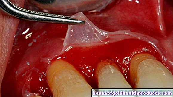
- Slit lamp examination: As part of this microscopic examination, the foreground of the eye is examined more closely. In the case of anterior uveitis, inflammatory cell material up to pus (hypopyon) and proteins (Tyndall phenomenon) can be seen in the anterior chamber of the eye (between cornea and iris).
- Reflection of the fundus of the eye: This examination, also known as funduscopy (ophthalmoscopy), primarily helps to diagnose middle and posterior uveitis. For a better view of the fundus, the doctor will give the patient eye drops to enlarge the pupil before the start of the examination.
- Eyesight test (using an eye test)
- Measurement of intraocular pressure (tonometry): This allows glaucoma to be recognized early as a possible complication of uveitis.
- Fluorescence angiography: This is a representation of the retinal vessels using a fluorescent dye. In this way it can be determined whether the place of sharpest vision on the retina (macula) is affected by the inflammation.
If there is a suspicion that uveitis is a side effect of another disease, further examinations are carried out to clarify. For example, blood tests and swabs from the conjunctiva help to identify an infection with bacteria, viruses or other pathogens as a cause of the eye inflammation.
Blood tests and imaging procedures (x-rays, magnetic resonance imaging, etc.) can provide information about various rheumatic or inflammatory diseases. For example, if sarcoid is suspected, a chest x-ray (chest x-ray) is usually very informative.
Exclusion of other diseases
Some conditions cause symptoms similar to uveitis. The doctor excludes these differential diagnoses in his examinations. These include, for example:
- bacterial or viral inflammation of the cornea (keratitis)
- pure retinal inflammation (retinitis)
- Episcleritis (inflammation of the connective tissue between the dermis and the conjunctiva)
- Tenonitis (a special form of dermis inflammation)
- certain types of glaucoma (angle closure glaucoma, hemorrhagic glaucoma)
Uveitis: treatment
Uveitis therapy depends on the cause of the eye infection.
In the case of non-infectious uveitis, which is not caused by pathogens, the doctor usually treats with corticosteroids ("cortisone"). They have an anti-inflammatory effect and are usually used in the form of eye drops, sometimes also as an eye ointment. If necessary, the doctor may also prescribe non-steroidal anti-inflammatory and pain relievers (NSAIDs).
Especially in severe cases of uveitis, the cortisone has to be taken as a tablet or injected into or around the eye. Other immunosuppressants such as azathioprine or cyclosporine may also be used.
So that the iris does not stick to the lens, the doctor also prescribes pupil-dilating eye drops (mydriatics such as atropine or scopolamine) for anterior uveitis.
In the case of infection-related uveitis, those affected receive medication against the causative pathogens if possible, for example antibiotic eye drops (possibly also antibiotic tablets) for bacterial uveitis or antivirals for viral eye skin inflammation.
In some cases, further therapeutic measures are necessary, such as surgery or other medication. If uveitis occurs, for example, as part of a rheumatic disease (such as reactive arthritis, juvenile idiopathic arthritis, etc.), it must be treated appropriately - for example with rheumatoid drugs such as methotrexate. If the intraocular pressure is increased, doctors also lower it with medication or by means of an operation.