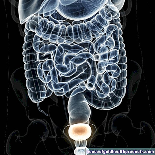
uterus
Eva Rudolf-Müller is a freelance writer in the medical team. She studied human medicine and newspaper sciences and has repeatedly worked in both areas - as a doctor in the clinic, as a reviewer, and as a medical journalist for various specialist journals. She is currently working in online journalism, where a wide range of medicine is offered to everyone.
More about the experts All content is checked by medical journalists.The womb (uterus) is the fruit holder or brood chamber in which the embryo develops until it is born. The contractions of the muscular wall of the uterus during birth (labor) give birth to the child. Read everything you need to know about: How is the uterus structured? Where is the uterus located? What is her job? What health problems can affect the uterus?
What is the womb?
The uterus is a muscular organ in the shape of an upside-down pear. Inside the uterus is the uterine cavity (Cavum uteri) with a flat, triangular interior. The upper two thirds of the uterus is called the body of the uterus (corpus uteri) with the dome (fundus uteri) in the uppermost area, which protrudes over the exit of one fallopian tube on the right and left. The lower, narrow third is called the cervix (cervix uteri).
Between the corpus uteri and the cervix there is a narrow connecting piece (isthmus uteri) that is about half a centimeter to a full centimeter long. This area is anatomically part of the cervix, but its interior is lined with the same mucous membrane as the corpus uteri. However, the mucous membrane in the isthmus - in contrast to that in the body of the uterus - does not take part in the cyclical changes in the context of the menstrual cycle.
The inner cervix forms the transition between the isthmus uteri and the cervix. The lowest part of the cervix that protrudes into the vagina is called the portio vaginalis uteri. In its center, the cervix joins the external cervix.
The uterus is usually slightly bent forward (anteversion) and bent slightly forward relative to the cervix (anteflexion). It rests on the bladder like this. Depending on the filling of the urinary bladder, the uterus shifts a little.
Uterus size and weight
The size of the uterus in an adult, non-pregnant woman is about seven to ten centimeters. The uterus is one and a half to three centimeters thick and weighs about 50 to 60 grams, which can increase to about one kilogram during pregnancy.
Structure of the uterine wall
The wall structure in the uterus shows three layers: The outer layer is a lining with peritoneum, the connective tissue perimetrium. Inwardly, there is a thick layer of muscle cells called the myometrium. There is a mucous membrane on the very inside. In the area of the uterine cavity, this is called the endometrium. It differs in its structure from the mucous membrane in the cervix.
What is the function of the uterus?
The function of the uterus only comes into play during pregnancy: the uterus provides the space in which the fertilized egg cell develops into a viable child.
The uterus prepares itself for this task every month: the endometrium thickens in the first half of the cycle under the influence of hormones (estrogens) to a thickness of about six millimeters. In a further step, the hormone progesterone unfolds its effect: It prepares the endometrium for the implantation of a potentially fertilized egg cell. If fertilization has not taken place, the thickened mucous membrane is shed and excreted via the menstrual period (the blood from torn mucous membrane vessels). The strong muscle layer inside the uterus contracts in order to transport the rejected tissue to the outside. These muscle contractions can be perceived as menstrual pain to different degrees.
If, however, an egg cell is fertilized within a cycle, it will implant itself in the lining of the uterus. This continues to grow to ensure that the embryo is nourished. The uterus can adapt to the child's growth and can reach an internal volume of up to five liters.
Where is the uterus located?
The uterus is located in the woman's pelvis, between the bladder and rectum. The perimetrium extends from the upper end to the front surface of the uterus, which rests on the urinary bladder, and further down to the isthmus, where it continues onto the urinary bladder. In the back of the uterus, the perimetrium rests on the uterus down to the cervix.
The uterus is held in place by various connective tissue structures (straps). In addition, the pelvic floor muscles usually prevent the uterus from sagging.
What problems can the uterus cause?
In 10 to 20 percent of women, the uterus is inclined backwards (retroversion) and / or bent backwards (retroflexion). This can be congenital, based on different filling states of the neighboring organs (such as the urinary bladder) or caused by inflammation or tumors. Some of the women affected suffer from symptoms such as back pain, pain during sexual intercourse, particularly severe, persistent menstrual pain (dysmenorrhea), increased menstrual bleeding (hypermenorrhea) or even sterility.
In endometriosis, the lining of the uterus (endometrium) also grows outside the uterus, for example in the fallopian tubes, in the ovaries, in the vagina, in the peritoneum or - albeit rarely - in regions outside the genital area, for example in the groin, in the rectum , in lymph nodes, in the lungs, or even in the brain. These endometrial foci also take part in the menstrual cycle, i.e. they are built up and broken down cyclically (including a small bleeding that is absorbed by the surrounding tissue). Common symptoms of endometriosis include abdominal pain, cyclical back pain, painful sex, menstrual irregularities, and infertility.
There are various malformations that affect the uterus. In the uterus bicornis unicollis, for example, there are two uterine bodies with a common cervix (cervix). If there are two bodies of the uterus, each with its own cervix, one speaks of uterus bicornis bicollis. Women with a didelphus uterus have two uteri of the same size, two cervices and usually two sheaths. If there is no uterus from birth, doctors speak of uterine aplasia.
The uterus can lower itself (i.e. go deeper into the pelvis), usually together with the vagina. Because of the tight connective tissue connections, the neighboring organs, urinary bladder and / or rectum, are also taken along. This lowering (descent) of the pelvic organs is a progressive process. Ultimately, the uterus can partially or completely emerge from the vagina (prolapse). Risk factors for the descent of the pelvic organs are weakness or injury to the pelvic floor (such as birth injuries), obesity, chronic cough and chronic constipation.
A cancer in the area of the cervix is called cervical cancer (cervical cancer). Risk factors include early first sexual intercourse, frequently changing sexual partners and poor genital hygiene. These factors increase the risk of human papillomavirus (HPV) infection. These germs are involved in the development of cervical cancer.
More often than in the cervix, a malignant tumor develops in the area of the corpus uteri; then there is uterine cancer (corpus cancer). Risk factors are, for example, old age, obesity (very overweight), diabetes mellitus and high blood pressure. Women who have never had a child are also more prone to uterine cancer.
Uterine polyps are caused by estrogen-induced hyperplasia (enlargement / increased growth) of the endometrial tissue. Uterine fibroids are benign muscle growths in or on the uterus, the growth of which is determined by estrogen. Both polyps and fibroids can cause discomfort, but do not have to be.
Tags: baby toddler dental care foot care
