heart
and Florian Tiefenböck, doctorEva Rudolf-Müller is a freelance writer in the medical team. She studied human medicine and newspaper sciences and has repeatedly worked in both areas - as a doctor in the clinic, as a reviewer, and as a medical journalist for various specialist journals.She is currently working in online journalism, where a wide range of medicine is offered to everyone.
More about the expertsFlorian Tiefenböck studied human medicine at the LMU Munich. In March 2014, he joined as a student and has supported the editorial team with medical articles ever since. After receiving his medical license and practical work in internal medicine at the University Hospital Augsburg, he has been a permanent member of the team since December 2019 and, among other things, ensures the medical quality of the tools.
More posts by Florian Tiefenböck All content is checked by medical journalists.
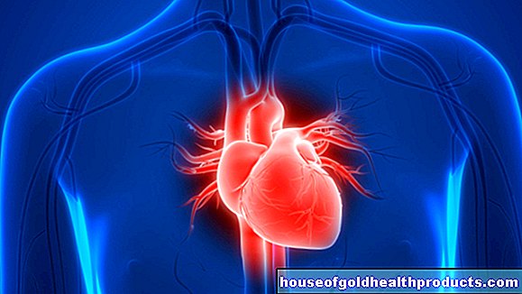
The human heart is a strong, cone-shaped hollow muscle. It lies in the rib cage behind the breastbone with the point towards the left. As the motor of our blood circulation, it is extremely powerful - it pumps around 8,000 liters of blood through the body every day. When exercising, it can increase its performance up to five times and more. Read everything you need to know about the structure and location of the heart, its function and important diseases!
What is the heart
The human heart is a strong, cone-shaped hollow muscle with a rounded tip. The heart muscle in an adult is about the size of a fist and weighs an average of 250-300 grams, and a woman's heart is usually slightly lighter than that of a man. Incidentally, the critical heart weight starts at around 500 grams. Heavier hearts can hardly be properly supplied with blood and supplied with enough oxygen. There is a risk of a heart attack.
Atria and ventricles
The structure of the heart is adapted to the complex function of the organ as the motor of the blood circulation. The cardiac septum divides the hollow muscle into a left and a right half, each of which is divided into two compartments: the left and right atrium (atrium) and the left and right heart chamber (ventricle).
From the outside you can see the subdivision into atria and heart chambers on the coronary furrow, a ring-shaped depression (sulcus coronarius). From here further heart furrows run towards the apex of the heart. The so-called sulci interventriculares can be recognized from the outside as the cardiac septum inside. Coronary vessels run in the coronary furrows, which doctors also call coronary arteries, coronary arteries or coronary vessels.
Auricular ears
The heart also has two so-called cardiac ears, a right and a left. They insist on muscle tissue. The right atrial appendage is located next to the main artery, the left on the pulmonary artery. Researchers do not yet know exactly what function the auricles play. What is clear, however, is that they form an important protein, the ANP (atrial natriuretic peptide). This messenger substance regulates the salt and water balance and in this way also the blood pressure. In addition, the auricular ears cover niches that arise between the outgoing vessels and the base of the heart.
Heart skeleton
The heart consists mostly of muscle tissue. But it also has a framework of tight connective tissue, the so-called heart skeleton. Not only the muscles are attached to it. It also forms rings of connective tissue fibers on which the heart valves sit. The heart skeleton thus plays a crucial role in the functioning of the heart.
heart valves
Because the heart valves are crucial for the blood to flow in certain directions in the heart: the atrium and ventricle on each side are connected via a heart valve. The mitral valve is located between the left atrium and the left ventricle, the tricuspid valve between the right atrium and the right ventricle. They prevent the blood from flowing back into the atria.
At the upper end of the heart muscle, the heart base, the large vessels branch off: from the right ventricle the pulmonary artery (arteria pulmonalis), which supplies the pulmonary circulation (small blood circulation). The pulmonary valve is interposed here to ensure that the blood does not flow back into the right ventricle. The main artery (aorta), which supplies the body's circulation (large blood circulation), branches off from the left ventricle. The aortic valve is located between the left ventricle and the aorta to prevent blood from flowing back into the left ventricle.
Heart wall layers
In the exact anatomy of the heart, the heart wall consists of three layers. From the outside in, these are:
- Epicardium (outer skin of the heart, part of the pericardium)
- Myocardium
- Endocardium
The endocardium consists of endothelial cells and connective tissue. The heart valves also emerge from the inner lining of the heart. The myocardium is the actual heart muscle. The outer skin of the heart, the epicardium, is a single layer of cells. There is also connective and fatty tissue there. This is where the nerves and blood vessels of the heart branch. The epicardium is also the inner leaf of the pericardium.
Coronary arteries
The muscles are supplied with all important nutrients and oxygen via special blood vessels. You can read more about these coronary vessels here.
Pericardium
A thin covering, the epicardium, rests on the myocardium. In addition, it is embedded in a sack made of connective tissue. You can read more about this pericardium in our article Pericardium
What is the function of the heart?
The function of the heart is to move the blood in the circulatory system, more precisely in the small and large bloodstream. The body motor acts like a pressure and suction pump in which valves - the various flaps - regulate the direction of the current (blood flow). They ensure that the blood is always pumped in the right direction and does not flow back.
Electrical impulses are necessary so that a human heart can contract (contract) regularly and in an orderly manner and allow the blood to flow in the vessels. For this there is a "pacemaker" (sinus node) in the right atrium. It independently generates electrical impulses that are distributed over the atria and stimulate the muscle tissue to contract. Via the AV node, a switching point between the atria and the ventricles, the signal reaches the ventricles, which then also contract - the heart "pumps". These excitation waves can be visualized in the EKG (electrocardiogram).
The heart skeleton also plays an important role in this conduction system. It is used for electrical insulation between the atria and the heart chambers. As a result, the electrical impulses do not spread indiscriminately from the atria of the heart to the entire ventricular musculature.
Myocardium - strong muscles
You can read more about heart muscle cells and their function in our article Myocardium
Where is the heart
The human heart lies in the rib cage behind the breastbone. Its broad base points to the top and back to the right, its rounded tip to the bottom and left to the front. The right half of the heart lies on the anterior chest wall. The left heart turns more to the left and backwards. The position of the heart in the chest offers special protection. Because behind the heart lies the spine. The ribs and sternum protect the heart at the sides and in front.
Where is the heart Which neighboring organs does it have?
The heart organ is surrounded by a bag of connective tissue, the pericardium, and is located in the lower middle mediastinum. This is the chest space between the two lungs. They are to the right and left of the heart. The heart lies below the diaphragm. Above the base of the heart, where the vessels branch off, the windpipe (trachea) divides into the two main airway branches, the bronchi. The main artery here runs in an arc over the left bronchus. The left atrium touches the esophagus posteriorly.
Which side is the heart on?
The right edge of the heart is about a good thumb's width to the right of the breastbone, about at the level of the second to fourth rib. To the left, the heart stretches diagonally downwards to the apex of the heart. It lies roughly between the fifth and sixth rib on an imaginary line that goes straight down from the middle of the collarbone. This means that around two thirds of the heart is in the left half of the chest.
What problems can the heart cause?
In people with heart failure (heart failure), the body motor is not able to generate the necessary pumping power. The disease can be acute or chronic. In severe cases, doctors have to replace the heart organ with a donor organ (heart transplant).
If the hollow muscle does not contract properly, there are arrhythmias. The most common forms include atrial fibrillation and atrial flutter. If people have a very slow heartbeat, doctors call it bradycardia. The opposite is the racing heart, medically called tachycardia.
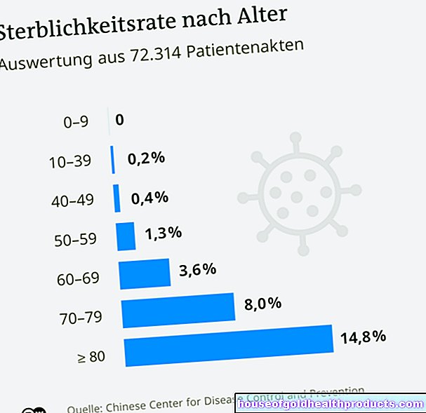
Coronary arteries constricted by fat and other deposits (atherosclerosis or arteriosclerosis) can only insufficiently supply the body motor with oxygen and nutrients. Doctors speak of coronary heart disease (CHD). It is the leading cause of death in the western industrialized countries. Because in the worst case, CHD can lead to a heart attack (myocardial infarction).
The heart valves can leak from birth or leak in the course of life. If the heart valve has a serious defect, they will no longer close or open properly. As a result, the blood flows back into the atrium or chamber or is no longer transported properly. Sometimes those affected then need an artificial valve.
Holes in the heart septum (septal defect) are among the most common congenital heart defects. More rarely, they are acquired in the course of life. They are mainly found in the ventricular septum, and more rarely in the atrial septum. Such a "hole in the heart" allows blood to flow from the left heart directly into the right heart.
In addition, various pathogens can attack the heart. As part of a viral or bacterial infection, there is a risk of heart muscle inflammation (myocarditis), especially if the sick do not take physical care, or inflammation of the inner lining of the heart (endocarditis). Patients with artificial heart valves or severe heart defects are particularly at risk from this.
Tags: Diagnosis fitness digital health