Arthroscopy
Valeria Dahm is a freelance writer in the medical department. She studied medicine at the Technical University of Munich. It is particularly important to her to give the curious reader an insight into the exciting subject area of medicine and at the same time to maintain the content.
More about the experts All content is checked by medical journalists.With the help of arthroscopy or joint endoscopy, larger joints in particular can be examined and damage to the joint structures treated. For this purpose, a special optical probe (arthroscope) is inserted into the joint through a small incision in the skin. Read all about arthroscopy, how it is done and what the risks are.

What is an arthroscopy?
Arthroscopy is a form of examining a joint. To do this, a so-called arthroscope is inserted through a small incision in the skin. This is a thin tube with a video camera at the end. A light source and a rinsing and suction device are also installed so that the doctor can fully observe the joint structures. In addition, special instruments can be used arthroscopically so that damage and injuries can be treated as soon as they are diagnosed.
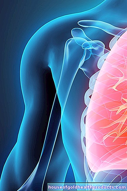
Arthroscopy of the shoulder
The shoulder is a particularly complex and sensitive joint that is often examined arthroscopically. You can read more in the article Arthroscopy - Shoulder.
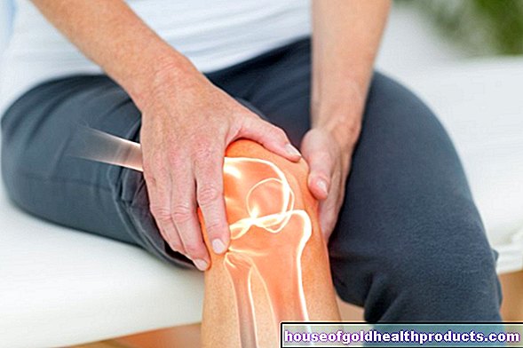
Arthroscopy of the knee joint
Injuries to the knee joint are particularly common. They can often be diagnosed and treated with an arthroscopy. You can read more in the article knee mirroring.
When do you do an arthroscopy?
Arthroscopy is primarily used to clarify joint complaints and examine joint injuries. The most common reasons are:
- Injuries or changes caused by an accident (traumatic)
- degenerative changes (joint wear) such as osteoarthritis
- inflammatory changes
As part of an arthroscopy, the doctor can often perform necessary operations with the help of additional instruments, which are usually introduced into the joint through additional skin incisions. This procedure is also known as minimally invasive surgery (MIS) or keyhole surgery.
Compared to the open surgical procedure, it has the advantage that healthy joint structures are spared and the organism is less stressed, the pain after the operation is lower and the healing time is usually shortened. The most common indications for arthroscopy include:
- Cartilage and bone damage
- Tears in ligaments, tendons, and muscles
- Bursitis
- free joint bodies
What do you do with an arthroscopy?
Before the actual arthroscopy, the patient's history is asked (anamnesis) and the patient is informed about the benefits and risks of the examination. In addition, a blood test is carried out, for example to detect a reduced ability of the blood to clot.
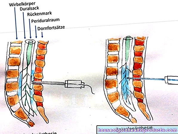
Arthroscopy is performed either under general anesthesia or under regional anesthesia, for which only the surgical area or one extremity is anesthetized. To prevent blood clots from forming during and after the examination, the patient is injected with an anticoagulant drug (heparin).
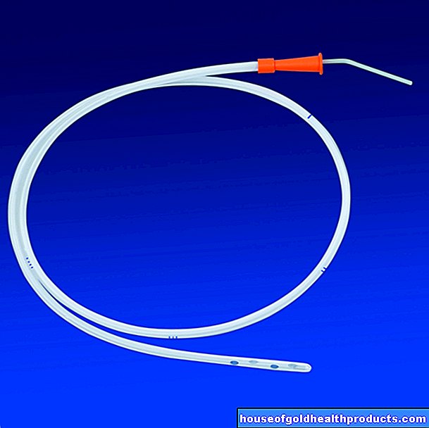
The skin of the operating area is depilated and carefully disinfected. Now the surgeon opens the joint through a small incision in the skin, through which a guide tube (trocar) is inserted. Sometimes it is necessary to fill the joint cavity with a sterile liquid or carbon monoxide and to stretch it so that the doctor can better orient himself in the joint space and clearly distinguish the joint structures.
The doctor finally inserts the arthroscope through the trocar. He follows the camera recordings in real time on a screen so that he can examine the joint while it is moving. If he discovers joint damage that can be treated arthroscopically, he introduces additional instruments into the joint cavity through additional skin incisions. With the help of burrs, for example, bones and cartilage can be smoothed, needle and thread allow the suturing of torn ligaments.
Finally, the arthroscope and all other instruments are removed and the skin incisions carefully sutured. If there is a risk of bleeding, drainage tubes can be temporarily inserted into the joint. A slightly compressing bandage also prevents bruising and protects the wounds from infection.
What are the risks of an arthroscopy?
Arthroscopy is a relatively uncomplicated examination. However, in rare cases the instruments and arthroscope used can injure the joint and joint structures such as cartilage and ligaments. As with any invasive procedure, structures such as vessels and nerves can also be damaged. In addition, bruises (hematomas) and bleeding may occur.
Infection of the wounds or the joint cavity can also occur. However, this complication is much less common with arthroscopy than with open surgical procedures. Despite anticoagulant medication, there is a risk of blood clots forming in the veins (thrombosis), as after any operation.
What do I have to consider after an arthroscopy?
Both after an outpatient arthroscopy and after an arthroscopy performed in a hospital, the affected joint should be kept as still as possible for a certain period of time. Pain reliever and inflammation-relieving medication, cooling and decongestant measures such as elevation are also used to guarantee quick and best possible healing.
After this phase of immobilization, an arthroscopy is followed by physiotherapy, which restores the function of the joint.
Tags: drugs desire to have children symptoms