Bone scintigraphy
Dr. med. Philipp Nicol is a freelance writer for the medical editorial team.
More about the experts All content is checked by medical journalists.Bone scintigraphy (also known as skeletal scintigraphy) is a nuclear medical examination with which bones and bone metabolism can be examined. Various diseases and injuries can be diagnosed, for example rheumatism, tumors and broken bones. Read everything you need to know about bone scintigraphy here!

What is a bone scintigraphy?
Bone scintigraphy is a sub-form of scintigraphy. Bones and their metabolism can be assessed very well with it. For this purpose, the patient is injected with a radioactively marked substance (radionuclide) via a vein, which is preferentially deposited in the bone, the stronger the higher the local metabolic activity. The radiation emanating from the radionuclide can then be measured and displayed as an image. In bone scintigraphy, phosphonates labeled with technetium-99m (99mTc) are used for radioactive labeling.
In some cases it is sufficient to examine individual bones or individual sections of the skeleton by scintigraphy (partial-body skeleton scintigraphy). This reduces the radiation exposure for the entire organism. In other cases, a full-body skeleton scintigraphy is necessary, for example to detect daughter tumors (metastases) of cancer tumors (for example breast, prostate, lung or kidney tumors).
In the so-called multi-phase scintigraphy (3-phase skeletal scintigraphy), recordings are made at different times (phases). This allows the blood flow in the arteries and veins to be shown. This is a good way of assessing inflammatory processes in bones or bone metastases, for example.
When to do a bone scintigraphy
Many diseases and injuries to the bones are associated with increased or decreased metabolic activity and can therefore be easily recognized with a scintigraphy.
For example, an increased metabolism can be measured in the area of a broken bone. Bone scintigraphy can therefore be used to depict broken bones, even if normal X-rays are sufficient in most cases.
A bone scintigraphy can also be used to diagnose various diseases such as:
- Bone cancer and bone metastases
- Inflammation of the bones (osteomyelitis, spondylodiscitis)
- Rheumatoid arthritis ("rheumatism")
- Bone infarction
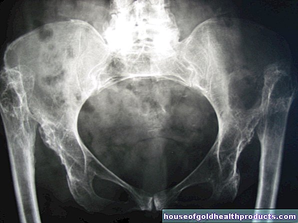
- Bone metabolism disorders such as Paget's disease or osteomalacia (painful softening of the bones)
In addition, unclear bone and joint complaints as well as complaints with joint prostheses (loosening, inflammation) are often clarified using bone scintigraphy.
Bone Scintigraphy: Risks
As with all scintigraphy methods, bone scintigraphy also has a certain level of radiation exposure, but this is only minimal. For an adult patient, it corresponds approximately to one to three times the annual natural radiation exposure in Germany. Children receive a lower dose of radioactively labeled substance for the examination, which means that the radiation exposure of the bone scintigraphy is even lower for them.
Tags: diet desire to have children travel medicine
.jpg)