Temporal arteritis (giant cell arteritis)
Ricarda Schwarz studied medicine in Würzburg, where she also completed her doctorate. After a wide range of tasks in practical medical training (PJ) in Flensburg, Hamburg and New Zealand, she is now working in neuroradiology and radiology at the Tübingen University Hospital.
More about the experts All content is checked by medical journalists.Temporal arteritis is the most common manifestation of giant cell arteritis - a rheumatic disease in which blood vessels become inflamed. Those affected mainly suffer from one-sided, severe headaches in the temporal region. The disease is diagnosed using ultrasound and a tissue sample. Since it can have serious consequences such as blindness, it must be treated quickly. Here you can read everything you need to know about temporal arteritis.
ICD codes for this disease: ICD codes are internationally recognized codes for medical diagnoses. They can be found, for example, in doctor's letters or on certificates of incapacity for work. M31

Temporal arteritis: description
In temporal arteritis, the artery in the temporal region becomes inflamed. It is the most common type of giant cell arteritis. This is a rheumatic disease in which blood vessels become inflamed (vasculitis). Some use the term temporal arteritis to mean giant cell arteritis.The diseases are also called Horton's disease, Horton's temporal arteritis or cranial arteritis in some places. However, these terms are considered out of date.
In fact, temporal arteritis is a symptom of giant cell arteritis. In the context of this vasculitis, however, vessels other than those in the temporal region can also become inflamed. Temporal arteritis can also occur in other inflammatory diseases.
Giant cell arteritis
Large and medium-sized vessels are affected in this vasculitis. The disease occurs most frequently at branching off vessels in the carotid artery. These vessels supply the temporal region, the back of the head and the eyes with blood. In some patients, giant cell arteritis affects the main artery or larger vessels in the trunk and extremities. Coronary arteries can also be affected (coronaritis).
Giant cell arteritis and thus also temporal arteritis are autoimmune diseases. This is because certain cells of the immune system (granulocytes and lymphocytes) accumulate in the affected vessels and form a chronic inflammation. Under the microscope you can see particularly large cells, so-called giant cells. This also explains the official name of giant cell arteritis. The disease causes cells in the vascular wall to multiply and eventually narrow the affected vessel. As a result, the blood supply can no longer be sufficient, especially during physical exertion. Depending on the organ system, corresponding symptoms are produced.
frequency
Giant cell arteritis is one of the most common rheumatic vascular diseases and the most common vasculitis. It usually shows itself through temporal arteritis. The disease rate increases with age. Women are significantly more often affected by giant cell arteritis than men. About half of the sick also have polymyalgia (polymyalgia rheumatica). The distinction between temporal arteritis or giant cell arteritis and polymyalgia is often difficult.
In polymyalgia rheumatica, too, large arteries become inflamed, especially the collarbone artery. Doctors assume that polymyalgia rheumatica is a mild form of giant cell arteritis, but primarily affects joints and tendons. Therefore, those affected usually complain of severe shoulder and upper arm pain and often also ailments in the pelvic area.
Temporal arteritis: symptoms
Almost all patients with Horton's temporal arteritis have particularly severe headaches. Most of them, however, have general symptoms long before the first headache.
Headache with temporal arteritis
Over 70 percent of people with temporal arteritis suffer from new, severe headaches. These are usually described as piercing to piercing and usually occur on one side of the temple. The pain increases when people chew, cough, or turn their heads. This is because an artery is affected, which supplies the masticatory muscles with oxygen and nutrients. If those affected chew solid foods, the masseter is more stressed and needs more nutrients. If the supply of a damaged artery cannot be guaranteed, pain in the area of the temples, scalp or a painless feeling of the locked jaw occur (claudication masticatoria). In some cases, this means that those affected have to take a break during a meal.
Visual disturbances in giant cell arteritis of the eye vessels
If, in addition to or instead of temporal arteritis, inflamed vessels appear in the eye, both the optic nerve and the eye muscles may function to a limited extent. The optic nerve, like the muscles, needs a constant supply of blood. If the supplying arteries change abnormally, visual disturbances can occur. These include fleeting eye dropouts (amaurosis fugax), in which those affected suddenly cannot see anything in one eye. If only a part of the image fails, it is called a scotoma. Visual impressions may be perceived as flickering images. If the eye muscles are supplied with too little blood, double vision, pain when turning the gaze or a drooping eyelid can occur. In the worst case, those affected may go blind due to temporal arteritis.
Other symptoms of temporal arteritis and giant cell arteritis
Some time before the typical headache of temporal arteritis occurs, those affected often suffer from unspecific symptoms. You feel exhausted or have a slightly increased body temperature. If only the main artery is attacked in giant cell arteritis, fever can be the only symptom of the disease. In addition, a lack of appetite and weight loss are accompanying symptoms of giant cell arteritis.
In addition to temporal arteritis or inflammation of the eye vessels, the following symptoms can also occur in giant cell arteritis:
Central neurological failures: If internal vessels are also affected by Horton's disease, for example, brain regions cannot be adequately supplied with oxygen and nutrients - this can lead to a stroke with symptoms such as paralysis, speech disorders or dizziness.
Sensory and movement disorders: In principle, every nerve in the body can be impaired if the blood vessel supplying it is restricted. This can worsen the feeling of the skin or even individual muscle movements.
Differences in blood pressure and arm pain: If the main artery is affected, the blood pressure can differ between the two arms. In addition, a palpable pulse on the wrist disappears in some people. Other people affected suffer from pain in the arms, which mainly occurs during exercise (arm claudication).
Aneurysm and dissection: If it is a section of the main artery in the chest, bulges (aneurysm) and vascular tears (dissections) occur more frequently, which can be life-threatening.
Angina pectoris: Giant cell arteritis can also affect the coronary arteries (coronary arteries). With this coronaritis, sufferers feel similar symptoms as with a heart attack. These include, for example, a feeling of pressure and chest pain, a kind of oppression, racing heart, shortness of breath, sweating or dizziness.
In around 20 percent of cases, temporal arteritis occurs as part of polymyalgia rheumatica. Conversely, about 30 to 70 percent of patients with giant cell arteritis develop polymyalgia. Those affected then also suffer from pain in the shoulder, pelvic area or neck muscles. Unlike typical headaches, these pains are mostly symmetrical and do not develop quite as suddenly. In addition, there may be morning stiffness that improves as the day progresses. Depressive moods are also not uncommon.
Temporal arteritis: causes and risk factors
Temporal arteritis or giant cell arteritis is a rheumatic disease in which the immune system works incorrectly. Certain immune cells called T cells trigger an autoimmune reaction. Why this happens has not yet been adequately researched. The disease may be triggered by infections with viruses (chickenpox, rubella) or bacteria (Mycoplasma pneumoniae, chlamydia).
Since not all people with such infectious diseases develop temporal arteritis, for example, there is probably a genetic susceptibility. People with certain proteins on white blood cells (HLA-DR4) are more likely to develop this condition. Temporal arteritis is also seen more often in people with polymyalgia, another rheumatic pain disorder.
Temporal arteritis: examinations and diagnosis
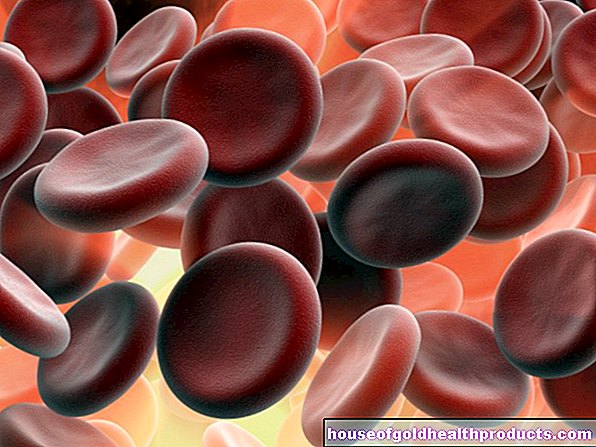
The right contact person for suspected temporal arteritis is a specialist in rheumatic diseases (rheumatologist) or for nervous diseases (neurologist). An American working group for rheumatological diseases (ACR) has put together various criteria that your doctor can use to diagnose temporal arteritis. To do this, he first conducts a consultation with a doctor (anamnesis) and, if there is a suspicion of illness, then carries out an imaging and tissue removal. A blood test could show increased levels of inflammation. If at least three of the following five criteria apply to an affected person, there is a probability of over 90 percent temporal arteritis:
- Age over 50 years
- First-time or novel headache
- Altered temporal arteries (painful tenderness, weakened pulse)
- Increased rate of sedimentation (blood test)
- Changes in the tissue of a temporal artery
Further investigations
In most cases, a specific ultrasound scan of the temporal arteries is done, in which the doctor can also show the blood flow (Doppler sonography). The temporal artery can also be assessed with magnetic resonance imaging (MRI). To do this, the person concerned is first injected with a certain contrast medium into a vein before his head is driven into the MRI tube on a movable couch. Vascular changes in other arteries, which can occur in giant cell arteritis, can also be shown under certain circumstances.
A restriction of the blood supply caused by the vascular inflammation can be examined more closely with positron emission tomography (PET). The examination process is similar to that of an MRI examination. A PET is mainly carried out if the main artery or other organ systems are affected, the patients suffer from pronounced accompanying symptoms or a tissue removal did not allow a clear diagnosis.
Tissue removal for temporal arteritis
If symptoms and imaging tests indicate temporal arteritis, in many cases a tissue sample (biopsy) is taken from the affected temporal region and examined under a microscope. Since the disease is not detected in an ultrasound examination in every patient, a tissue sample should be taken even if the ultrasound result is normal. In some cases, a piece of artery is removed from the other side of the temple.
The biopsy of the temporal artery is considered the gold standard for diagnosing temporal arteritis!
Before the biopsy, the doctor carefully selects the location of the sample collection. He also makes sure that the removed piece of vessel is sufficiently long (approx. One centimeter). This is because the inflammatory vascular changes with giant cells, as are typical for giant cell arteritis, only occur in sections in the vascular walls. The wall areas in between look normal.
Temporal arteritis: treatment
After temporal arteritis is diagnosed, the affected person should be treated immediately with a cortisone preparation. For the first four weeks, a dosage of one milligram of prednisolone per kilogram of body weight is recommended. If the symptoms have disappeared as a result of the therapy and the inflammation levels in the blood have normalized, the dose should be reduced continuously. If symptoms reappear, more prednisolone must be taken again. The attending physician works out a precise intake schedule with his patient. If blindness is imminent, prednisolone therapy must be administered in high doses via the vein for three to five days.
If giant cell arteritis affects the eye vessels, it is a medical emergency! Permanent blindness threatens!
Since long-term therapy with cortisone preparations can cause numerous undesirable side effects, further medication must be taken. Calcium and vitamin D reduce the risk of developing osteoporosis (fragile bones). A proton pump inhibitor protects the lining of the stomach. In addition, blood sugar should be checked regularly and treated if necessary. While experts used to recommend the preventive intake of the "blood thinner" ASA (acetylsalicylic acid), the hoped-for prophylactic effect has not been confirmed in the meantime.
Temporal arteritis: disease course and prognosis
Without therapy, around 30 percent of those affected go blind. However, with early diagnosis and subsequent therapy, the symptoms disappear permanently in almost all patients. Giant cell arteritis rarely occurs repeatedly or turns into chronic temporal arteritis, for example.
Tags: hair teeth gpp


.jpg)