Endosonography
All content is checked by medical journalists.Endosonography is an ultrasound examination in which the transducer is not pressed onto the skin from the outside, but inserted into the body. Read everything about endoscopic ultrasound, what variants there are and what risks they can involve!
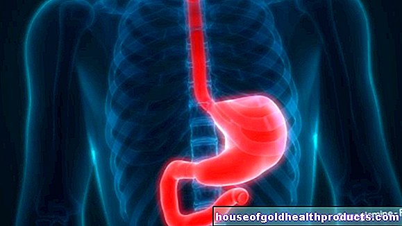
Endoscopic ultrasound of the stomach and esophagus (EGD)
An endosono of the stomach, esophagus, or duodenum can be done without anesthesia. On request, however, the patient can receive a calming agent so that he is relaxed and has no memories of the examination later. The doctor then inserts a flexible ultrasound probe into the stomach through the patient's mouth. With this he can look through the stomach wall through neighboring organs, for example the pancreas. He can also use endosonography to assess the wall layers of the esophagus and stomach and clarify whether there are tumors or inflammations, for example.
Endoscopic ultrasound of the airways (endobronchial ultrasound)
Endoscopic ultrasonography of the airways is similar to that of the gastrointestinal tract, the doctor simply guides the endoscope into the windpipe instead of the esophagus. In addition to the airways themselves, they can also assess the lymph nodes located near the windpipe or large bronchi, which is particularly important if cancer is suspected. If necessary, the doctor takes a tissue sample using a thin puncture needle under sonographic control.
Endobronchial ultrasound is a very safe procedure, but it can also cause injuries to the airways and bleeding when tissue is removed.
Transvaginal endosonography
During the transvaginal ultrasound examination, the gynecologist inserts a special thin, elongated transducer into the vagina. This enables him to assess the internal female sexual organs, i.e. the uterus, fallopian tubes and ovaries, particularly well. The first ultrasound during pregnancy is also carried out through the vagina.
The advantage of transvaginal endosonography over conventional ultrasound examinations through the abdominal wall is that it provides better images due to the proximity to the organs. In addition, the transvaginal ultrasound examination, like the conventional ultrasound scan of the abdominal wall, does not involve any particular risks and is only somewhat uncomfortable, but not painful for the patient.
Transrectal endosonography
The ultrasound examination of the rectum is primarily used to clarify fistulas, abscesses and tumors. The patient lies either on his side or on his back with his legs slightly bent and apart. The doctor inserts a thin, lubricated ultrasound probe into the patient's rectum. By carefully moving and tilting the transducer, the doctor can now assess the man's prostate gland in addition to the bowel itself. Endoscopic ultrasonography via the rectum is harmless, and slight pain in the anal region can sometimes occur.
Tags: home remedies stress symptoms


.jpg)