Atheroma
All content is checked by medical journalists.The atheroma is also known colloquially as a bag of groats. It is a benign cyst that is usually located in the scalp and protrudes into a hemispherical shape. Atheromas develop in the hair follicle and can swell up to the size of a chicken egg. Read more about causes and treatment of atheroma.
ICD codes for this disease: ICD codes are internationally recognized codes for medical diagnoses. They can be found, for example, in doctor's letters or on certificates of incapacity for work. L72
Atheroma: description
Doctors call atheroma a "bump" surrounded by a layer of skin that is mainly filled with skin cells and fat. Such filled cavities in the subcutaneous tissue, which develop due to a blocked gland, are also called retention cysts - in this case it is a trichilemmal cyst ("hair root sheath cyst"). Colloquially, the atheroma is also referred to as a groats bag.
Atheromas arise in the area of a hair root and therefore more often in areas of the body with a lot of hair. 90 percent of them are found on the hairy scalp, but sometimes also in other hairy areas.
Differentiation from the epidermoid cyst
Occasionally the so-called epidermoid cyst is also referred to as an atheroma. These pea- to plum-sized nodes develop from hair roots, but from their uppermost part (infundibulum). They mainly contain peeled horn material that is stacked on top of one another. The "real" atheroma, on the other hand, is primarily filled with a very fatty substance.
Atheroma: symptoms
An atheroma is an externally visible bump in the skin. It is full, but elastic rather than hard. Some atheromas can be moved under the skin. After an inflammation, however, they can also sit firmly on the scalp - 90 percent of all atheromas are found here. However, they can also occur on other parts of the body, such as the neck, chest, stomach or in the genital area. If several atheromas are present at the same time, they are sometimes grouped together.
Atheromas are usually one to two centimeters in diameter. However, they can swell up to the size of a chicken egg - in rare cases even the size of a tennis ball. With larger cysts, the skin that spans them is greatly stretched apart. This leads to the hair that grows here being further apart or missing entirely. In some cases, a gray or black point can be seen on the surface of the atheroma.
Usually, atheroma is the same color as the skin around it. It is primarily noticeable because of its bulge. Unless it becomes infected, it is painless and hardly causes any discomfort. Depending on the seat and size, however, those affected find it cosmetically annoying.
In the case of inflammation, the skin around the atheroma will be reddened, swollen, and touched or gently pressure will cause pain. If additional pus collects inside the atheroma capsule, it is an abscess.
Atheroma: causes and risk factors
An atheroma usually develops in the scalp from a hair root (follicle) - more precisely from the narrow canal in which the area of the hair that is still hidden under the skin is located. A small sebum gland opens into this canal in every hair. It ensures that the hair is coated with a film of oily liquid (sebum). If the sebum glands are very active, the hair quickly becomes greasy.
The duct of the sebum gland can be blocked in a certain area, the so-called isthmus, for example by small fat crystals or skin cells. The sebum can then no longer flow off unhindered, but the gland continues to produce it. Gradually, the sebum builds up and the hair root is pumped up into a round "bubble" - an atheroma develops.
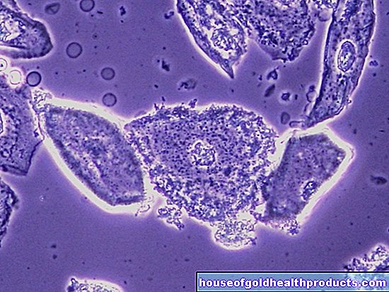
This mechanism also explains a special characteristic feature of atheroma: the structure of the skin that surrounds the cyst is similar to that of the skin that surrounds each individual hair root. One also speaks of a trichilemmal cyst. If you look at an atheroma cover under the microscope, you can usually see that it is keratinized with keratin. This hard material also forms hair and nails.This skin distinguishes the atheroma from other cysts and tumors.
Atheroma: examinations and diagnosis
The diagnosis of atheroma is usually made by a general practitioner or a dermatologist (dermatologist). In the first interview to collect the medical history (anamnesis) he asks the person concerned, for example, how long the cyst has existed, whether it causes him pain and whether there are or have been any other "bumps".
During the physical examination, the doctor takes a closer look at the "bump" - usually he can assess very quickly whether it could be an atheroma. He also checks how the cyst reacts to pressure and whether it can be moved.
Whether there is really a "real" atheroma (trichilemmal cyst) or an epidermoid cyst can sometimes only be determined with certainty after the "bump" has been surgically removed and histologically examined in the laboratory. A histological examination is also important to clarify whether it is perhaps a malignant growth.
Atheroma: treatment
If the atheroma is small, has stopped growing, and does not bother the person, sometimes treatment is not necessary. But if you have an atheroma that bothers you or continues to grow, you can have your dermatologist surgically remove it. Such an intervention is highly recommended if the atheroma is infected and inflamed, or if it is unclear whether malignant cells are involved.
Remove atheroma
The dermatologist usually removes an atheroma on an outpatient basis and under local anesthesia. During the procedure, the doctor makes sure that he cuts away the atheroma along with its capsule and the associated duct. If parts of it remain in the skin, there is a high risk that the atheroma will come back.
Under no circumstances should you remove an atheroma yourself, for example by trying to express its contents - manipulation with fingers and fingernails can introduce bacteria into the skin tumor. As a result of such a bacterial infection, the atheroma becomes inflamed.
When the atheroma becomes infected
With a bacterial infection, the atheroma swells, reddens, feels warm, and hurts to the touch. If pus accumulates within the cyst and cannot drain away, an abscess develops. In any case, this requires medical treatment. The doctor often uses an antibiotic for therapy.
Atheroma: disease course and prognosis
How fast an atheroma grows varies from case to case. Some atheromas only grow to a certain size and then stagnate.
In principle, after the surgical removal of an atheroma, an atheroma can develop again in the same place. But if the procedure has been carried out properly, the risk of it is low.
Tags: drugs desire to have children pregnancy birth


.jpg)