fracture
Dr. med. Mira Seidel is a freelance writer for the medical team.
More about the experts All content is checked by medical journalists.In the case of a fracture (broken bone), the continuity of the bone is completely or partially interrupted. This is usually associated with symptoms such as pain and loss of function. The cause of the break can be direct or indirect violence, a previous illness or fatigue (excessive stress). As a complication of a broken bone, the compartment syndrome is a surgical emergency. Find out more about fractures here.
ICD codes for this disease: ICD codes are internationally recognized codes for medical diagnoses. They can be found, for example, in doctor's letters or on certificates of incapacity for work. S62S22T08S12S32S02S82S92S42T02S72S52
Brief overview
- What is a fracture? Fracture is the medical term for a broken bone.
- Forms of fractures: e.g. open fracture (bone fragments are exposed), closed fracture (no visible bone fragments), dislocation fracture (fracture close to the joint with dislocation of the joint), spiral fracture (spiral-shaped fracture line).
- Symptoms: pain, swelling, restricted mobility, possibly misalignment, visible bone fragments with an open fracture
- First aid: immobilize the injured part of the body and stabilize it (e.g. with a triangular cloth in the event of a broken arm), if necessary elevate it, carefully cool the closed fracture (e.g. with an ice pack wrapped in fabric), cover the open fracture with sterile cover, call for rescue
- Treatment: either conservative (e.g. using a plaster cast) or surgical
- Prognosis: among other things, it depends on the location, type and severity of the fracture, the age and general health of the patient. With prompt, adequate therapy, a fracture usually heals well and without consequences.
Fraktur: description
Doctors understand a fracture as a broken bone: The bone is split into two or more fragments, which can also be displaced. This happens when the bones are subjected to direct or indirect force from outside, such as an accident.
Structure of bones
Humans have a total of 206 different bones. In some places, bones have "predetermined breaking points", for example on the upper arm, which is particularly prone to breakage. Every bone consists of mineral, elastic and connective tissue components. Blood vessels also run through the bone. Nerve fibers also run in the periosteum. Depending on the age of a person, the composition of his bones varies:
Children's bones are predominantly elastic. They therefore usually break as what is known as green wood fracture, in which the periosteum is still intact.
Adult bones have a balanced ratio of mineral, elastic and connective tissue components.
In older people, the bones lose elastic and connective tissue and therefore break more easily. In addition, due to the changed hormonal balance, the bones increasingly decalcify with age, which makes them brittle and fragile. A 70-year-old is therefore three times more likely to break a bone than a 20-year-old.
Fracture healing
Bone tissue heals without scars. The goal of bone fracture treatment is that the person affected can put weight back on the bone as soon as possible. Fast healing is achieved when the anatomical axis relationships of the bone are correct. In addition, the hernia should be immobilized and an adequate blood supply created.
The time it takes to heal a fracture varies depending on the section of the skeleton. For example, a broken collarbone only takes about three to four weeks with conservative treatment, while a broken thigh takes about ten to fourteen weeks to heal.
In children, a bone fracture heals faster because they are still growing and axial misalignments and shortenings can still be corrected. A broken bone in children can therefore usually be treated conservatively.
A broken bone can heal in two different ways and is treated differently depending on the type. Doctors differentiate between direct and indirect fracture healing.
Indirect fracture healing
Most often, the bone heals through indirect fracture healing. This means that the bone forms a so-called callus at the broken ends, a scar tissue of the bone that bridges the gap between the bone ends. Fracture healing takes place in five phases:
Injury phase: this is where the fracture happens.
Inflammatory phase: In the fracture zone, a bruise (hematoma) occurs, which is replaced over time by connective tissue cells such as granulocytes, mast cells and monocytes.
Granulation phase: In the next phase (four to six weeks) a soft callus of granulation tissue forms. The callus runs from the fracture ends towards the center. Since the ends of the fracture are poorly supplied with blood, bone necrosis (dead bone tissue) of a few millimeters occurs. The bone is therefore first slightly broken down in order to restore fragment contact. The dead bone tissue is broken down by so-called osteoclasts (bone-degrading cells), which is why you can see a widened fracture gap in the granulation phase in the X-ray image within the first two weeks. This is necessary for the bone to heal. Osteoblasts (bone-building cells) then replace the lost bone tissue with new ones.
Callus hardening phase: the connective tissue cells that migrate into the area of the hernia differentiate into cartilage-like cells under resting conditions. These mineralize slowly, which takes about three to four months. The new tissue becomes increasingly stronger. Special growth factors form a new substance in and on the outside of the fracture (braided bone). The person concerned notices this from the fact that the pain diminishes over time when the corresponding part of the body is moved.
Remodeling phase: The remodeling phase starts from the sixth month and can last up to a year. The still mesh-like new bone tissue is transformed into lamellar bones. In the X-ray this is shown by the beginning of new bone formation around the fracture. The initially unstructured, braided bone becomes more and more compact, which is supported by muscle tension. The initially spherical callus becomes flatter, so that after months or years the bony cortex is only insignificantly compressed.
Direct fracture healing
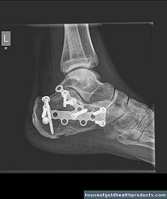
With direct fracture healing, the bone fracture heals without a visible callus. This is only possible with a bone fracture that fits directly on top of one another. Healing without a callus is therefore only possible through surgical measures. In the so-called compression osteosynthesis, the fracture is mechanically absolutely immobilized. This enables the ends of the bone to be adequately supplied with blood. In this way, new cells can develop on the fracture surface, which reproduce new bone tissue and link the fracture. No callus is therefore visible in the X-ray image. The previously visible fracture gap becomes blurred and at the end disappears completely.
Impaired fracture healing
A clearly prolonged fracture healing speaks for a disturbed fracture healing. In the X-ray you can see a widened fracture gap.
If no bony connection has formed at the two ends of the broken bone after four to six months, doctors speak of a "wrong joint" (pseudarthrosis).
Fracture: symptoms
Symptoms such as pain and swelling are typically associated with a broken bone. However, they are among the so-called uncertain signs of fracture, in contrast to misalignment of a limb as a visible sign of fracture.
Unsafe fracture signs:
- The movement can be carried out spontaneously.
- Pain on the move
- Loss of function of the joint
- swelling
Safe fracture signs:
- Misalignment
- wrong agility
- Crunching when moving
Open and closed fracture
If the skin over the fracture is open, it is an open fracture. It should first be covered in a sterile manner at the scene of the accident and only uncovered again under sterile conditions during the operation. This prevents germs from getting into the wound.
If the skin barrier over the fracture remains intact, it is a closed fracture. Sometimes nothing of the break can be seen from the outside. In other cases, abrasions through to extensive skin defects such as skin bruises are visible.
Fracture: examinations and diagnosis
The specialist responsible for suspecting a broken bone is a doctor specializing in orthopedics and trauma surgery.
anamnese
He will first ask you exactly how the accident happened and your medical history (anamnesis). Possible questions are:
- How did the accident come about? Was there any direct or indirect trauma?
- Where do you suspect a break?
- How do you describe the pain?
- Have there been previous injuries or previous damage?
- Have you had any complaints before?
Physical examinations
After the anamnesis discussion, the doctor examines the patient. He inspects the affected area looking for misalignments and swellings. He also feels whether it is tender or the muscles are particularly tense. He also checks whether the movement can be carried out correctly and whether it produces a creaking or grinding noise.
In the event of a (suspected) bone fracture, it is also important to check the peripheral blood flow, motor skills and sensitivity in the injured body region so as not to overlook any injured nerves, blood vessels or tendons! The distant pulses (e.g. on the foot with a broken leg) provide information about the blood circulation. To check motor skills, he asks you to actively move your fingers and toes. Sensitivity is also checked, for example when the doctor touches different areas of skin with a needle or finger.
Imaging
A subsequent x-ray examination in two planes can confirm the suspicion of a broken bone. If the pelvis or spine is affected, computed tomography (CT) is usually performed for a more detailed assessment. It can also be used to detect a so-called occult fracture - a broken bone that is not visible on X-rays.
The imaging shows where exactly the fracture gap runs and to what extent bone fragments are displaced. The break may have shifted sideways, shortened, lengthened, twisted or kinked in its axis.
Fracture: causes and risk factors
When it comes to the term fracture, most people think of a traumatic bone fracture: A sufficiently high force of force has broken the actually firm and elastic bone. However, a fracture can also result from an illness. There are basically three mechanisms that cause a fracture to develop:
- A direct fracture occurs when external force hits the healthy bones.
- A pathological fracture (spontaneous fracture) is usually the result of a pathologically altered bone such as tumor metastases, bone cysts or osteoporosis.
- A fatigue fracture (stress fracture) results from sustained mechanical stress, for example during long marches or running a marathon.
Fracture shapes
Depending on the violence and the shape of the bone, there are different types of fractures:
- Bending fracture: it is caused by direct or indirect impact on the bone. Tensile stress occurs on the concave side of the bone, which is why the bone there tears. On the other hand, on the convex side, the pressure is so great that a so-called flexible wedge is blown out. This happens, for example, with a direct impact on the shin.
- Torsional or torsional fracture: It is caused by indirect force, in which tensile stresses occur in the bone as a result of rotation. This break can occur, for example, when falling in a ski boot with a blocked safety binding.
- Spiral fracture: It has a spiral-shaped fracture gap and is caused by torsional loads. An axial load or gravity often also plays a role. Usually a spiral-shaped rotating wedge is created.
- Avulsion fracture: It can be caused by tensile forces acting on the bone via a ligament or a tendon attachment. The fracture line runs transversely to the direction of pull, for example in the case of an olecranon fracture (fracture of the upper edge of the ulna).
- Compression fracture (compression fracture): It usually occurs in the longitudinal axis of the body as a result of indirect force. This mostly affects the loose honeycomb structure of the cancellous bone, which is irreversibly compressed. Typical examples are the vertebral body fracture and the calcaneus fracture.
- Comminuted fracture: Here the bone is splintered into many (more than seven) fracture fragments due to a violent application of force. The bone fragments are typically displaced (dislocated). In addition, the surrounding soft tissues are severely injured. A classic example is a gunshot fracture or a comminuted fracture after a motorcycle accident.
- Dislocation fracture: This is a fracture near the joint in which the joint is also dislocated. There are two mechanisms of development: Either the dislocation is the cause of the fracture or the fracture and the dislocation have arisen at the same time. Dislocation fractures can occur in the ankle, tibia, or hip joint.
- Incomplete fracture: This refers to fissures and fissures that are not completely broken. One example is the child's greenwood fracture, in which the periosteum is still intact.
Fraktur: AO classification
The various bone fractures are classified by the AO, the working group for osteosynthesis issues. The AO classification is used to describe fractures exactly with a four-digit code, thus enabling treatment that is standardized worldwide. Relevant factors for the classification are:
- In which part of the body is the broken bone?
- At what point within this body region?
- Has the bone been stable?
- Are the bone fragments still supplied with blood?
- Is there any additional cartilage damage?
- Has the capsule-band apparatus been injured?
The AO classification is most often used for fractures on the long tubular bones such as the upper arm, forearm, thigh and lower leg bones. But hand and foot injuries, jaw fractures and fractures of the pelvis and spine can also be classified according to it.
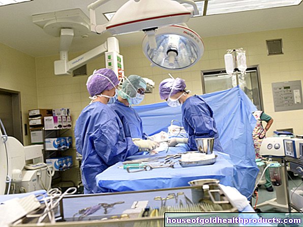
Fracture: treatment
How a fracture is treated depends primarily on the location, type and extent of the fracture and any accompanying injuries. In principle, the treatment can be conservative (e.g. with a plaster cast) or surgically.
You can find out how to properly provide first aid in the event of a broken bone and which therapy options are available to the doctor in the article Fracture: Treatment.
Fracture: disease course and prognosis
The prognosis for a fracture depends on both the type of injury and the appropriate treatment. The age and general health of the patient also have an influence.
In most cases, a fracture heals well and without consequences after adequate conservative treatment or surgery - but this often takes longer in older people than in younger people. In the case of open comminuted fractures and broken bones in which vessels have been affected, it is difficult to make an accurate prognosis. An infected fracture can result in the limb having to be amputated if sepsis ("blood poisoning") has developed. Long-lasting disorders often occur, particularly in the case of a joint fracture and fracture close to the joint.
Long lasting complications
Sometimes the broken ends do not grow back together in the bony but remain flexibly connected. Then a "wrong joint" developed - a pseudarthrosis. It manifests itself in swelling, overheating and pain when moving and exercising. There are the following causes for a pseudarthrosis:
- Movement in the fracture gap overloads the bone with the result that connective tissue tears and trabeculae break.
- Too much distance between the ends of the fracture prevents the ends of the fracture from touching and forming a bridge.
- If the soft tissues are damaged too much, they can reach into the fracture gap and lead to delayed healing.
- Smoking or non-cooperative behavior by the patient
Other long-lasting complications that can occur with a fracture are, for example, instabilities in the affected joint area, joint wear (osteoarthritis, arthrosis) and misalignments.
Tags: Diseases palliative medicine skin care