Seminoma
and Martina Feichter, medical editor and biologistFlorian Tiefenböck studied human medicine at the LMU Munich. In March 2014, he joined as a student and has supported the editorial team with medical articles ever since. After receiving his medical license and practical work in internal medicine at the University Hospital Augsburg, he has been a permanent member of the team since December 2019 and, among other things, ensures the medical quality of the tools.
More posts by Florian TiefenböckMartina Feichter studied biology with an elective subject pharmacy in Innsbruck and also immersed herself in the world of medicinal plants. From there it was not far to other medical topics that still captivate her to this day. She trained as a journalist at the Axel Springer Academy in Hamburg and has been working for since 2007 - first as an editor and since 2012 as a freelance writer.
More about the experts All content is checked by medical journalists.
Seminoma is the most common type of testicular cancer. It is generally operated on and - depending on the stage - further treated, for example with chemotherapy or radiation. Overall, the seminoma has a better prognosis than the other malignant testicular tumors. Find out everything you need to know about the seminome here.
ICD codes for this disease: ICD codes are internationally recognized codes for medical diagnoses. They can be found, for example, in doctor's letters or on certificates of incapacity for work. C62
Seminome: general
Seminoma is the most common type of testicular cancer. It is one of the so-called germ cell tumors (germinal tumors) and develops from the spermatogonia. These are precursors of the male germ cells (sperm). Other germ cell tumors of the testicle are grouped under the term non-seminoma. They arise from various other types of tissue.
Researchers assume that both seminomas and non-seminomas arise from the same preliminary stage - degenerate cells of embryonic development in the womb. This preliminary stage of testicular tumors is called testicular intraepithelial neoplasia (TIN). The very rare "spermatocytic seminoma" is an exception: It does not develop from the TIN, but directly from sperm-forming cells, i.e. only during the final sperm formation.
The World Health Organization also distinguishes other subtypes of the seminome. These include the classic seminoma and seminoma, which also contain cells from other types of tissue (such as connective and supporting tissue). But the latter are very rare. A pathologist can determine which type of testicular cancer is in each individual case by examining the removed tumor tissue.
The average age of seminoma patients is around 40 years.
Seminoma: symptoms
A palpable, painless induration in the scrotum is one of the most important signs of testicular cancer (like seminoma). Usually only one testicle is affected, more rarely both are pathologically changed.
An enlarged testicle can also be an indication of a testicular tumor. It is often accompanied by a feeling of heaviness. In addition, a pulling can occur that can radiate into the groin.
Another possible sign of testicular cancer (such as seminoma) is enlarged breasts, which can also be painful. Hormones produced by many testicular tumors are responsible for breast growth.
For more information about the signs of testicular cancer (such as seminoma), see Testicular Cancer Symptoms.
Seminoma: causes and diagnosis
It is not known exactly why some men develop seminoma (or another form of testicular cancer). However, we now know several risk factors that favor such a malignant tumor:
Accordingly, men who have had testicular cancer in the past are particularly at risk. An undescended testicle also increases the risk of a malignant testicular tumor - even if it has been surgically corrected. Genetic factors also seem to play a role in the development of seminomas (or testicular cancer). The same tumor occurs more frequently in some families. To read more about these and other risk factors for testicular cancer, see Testicular Cancer: Causes and Risk Factors.
How can a seminoma be diagnosed?
In a detailed discussion (anamnesis), the doctor asks the patient in detail about the symptoms (such as lumps in the testicles). He also asks about possible risk factors such as previous testicular cancer or undescended testicles. Patients should also report to their doctor about any testicular cancer in their close relatives.
This is followed by a physical exam. The doctor will palpate both testicles and the breast, among other things. A comprehensive blood test also provides important information. If, for example, the blood level of the protein AFP (alpha-fetoprotein) is increased, this can indicate testicular cancer - especially a so-called non-seminoma. In the case of a seminoma, on the other hand, the AFP value is normal.
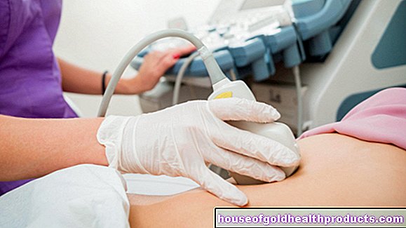
Imaging procedures such as computed tomography help determine the spread of the tumor.
You can read more about necessary examinations if a seminoma or testicular cancer is suspected under Testicular cancer: examinations and diagnosis.
Seminoma: treatment
As with other types of testicular cancer, surgery is the first step in treatment for a seminoma: the surgeon removes the diseased testicle, epididymis, and spermatic cord. This mandatory procedure is called ablatio testis or orchiectomy.
In a few cases it is possible not to remove the entire testicle but only the degenerated part. This procedure is particularly advisable for patients who only have one testicle. In this way, testosterone production, which takes place in the testicles, is still guaranteed.
Partial removal of the testicle can also be useful in another case: Testicular cancer usually only affects one testicle. To be on the safe side, however, one often takes a tissue sample from the second testicle and examines it for cancer cells. Only about five percent of testicular cancer patients will find what they are looking for - then bilateral testicular cancer is present. If possible, the surgeon leaves as much healthy testicular tissue as possible so that fertility and testosterone production are at least partially guaranteed. Sometimes, however, it is unavoidable to completely remove both testicles.
Further treatment after the operation depends on how far the tumor has progressed.
Seminoma: Stage I treatment
In stage I, the seminoma is limited to the testes. In examinations (such as computed tomography, CT) no lymph node involvement and no more distant cancerous settlements (distant metastases) could be detected. After the operation, the patient is generally considered cured. However, it cannot be ruled out with certainty that the tumor has not already formed the smallest metastases - so small that they cannot be detected using CT and other examinations. Depending on how great this possibility is, the operation is followed by one of three possible follow-up treatments: surveillance strategy, radiation therapy or chemotherapy.
1. Monitoring strategy
In Europe and the USA, the "wait and see" strategy is usually chosen for a seminoma in the early stages after the operation: the patient is thoroughly examined at regular intervals in order to detect a possible return of the cancer at an early stage.
2. Radiation therapy
For some seminoma patients (stage I), radiation therapy is recommended as a precautionary measure after the testicle has been removed: the back of the abdomen is irradiated. This is supposed to eliminate any existing tiny cancerous settlements in the lymph nodes along the abdominal artery. The irradiation takes place five days a week over a period of two weeks.
However, radiation therapy is only recommended in special cases for a stage I seminoma. After years or decades, the treatment itself can cause a malignant cancerous tumor (second tumor).
3. Chemotherapy
As an alternative to radiotherapy, chemotherapy can also be carried out as a precautionary measure after the testicle has been removed in seminomas (stage I). The patients receive a well-tolerated drug that can inhibit the multiplication of cancer cells (cytostatic). Chemotherapy is carried out once or twice. The patient does not have to stay in the hospital for this (outpatient chemotherapy).
Seminoma: treatment in stages IIA and IIB
In a seminoma in stage II, neighboring (regional) lymph nodes are affected by the cancer cells (more in IIB than in IIA). The patients then receive radiation therapy after the testicle is removed.
If irradiation is not possible for certain reasons, chemotherapy is chosen instead: In three cycles, the patient is given the three cytostatics (cytotoxic drugs, cell poisons) cisplatin, etoposide and bleomycin (PEB) in a vein.
Note: Clinical studies are currently investigating whether a seminoma in stage IIA or IIB can be treated more effectively with combined radiation and chemotherapy.
Seminoma: treatment in stages IIC and III
If the seminoma is even more advanced (stage IIC and higher), experts recommend three to four cycles of chemotherapy after the testicle has been removed. Here, too, the three cytostatics cisplatin, etoposide and bleomycin (PEB) are used.
Seminoma: disease course and prognosis
Seminoma has a relatively good prognosis, even in advanced stages - and is overall a better prognosis than the second main group of testicular cancer (non-seminomas). One reason for this is that the seminoma is less prone to the formation of daughter tumors (metastases) than a non-seminoma. Therefore, practically all patients with a seminoma stage I can be cured with the help of standard therapy. In stages IIA and IIB, the healing rate is over 95 percent. In higher seminoma stages (from IIC) 80 to 95 percent of patients can still be treated successfully.
In some cases, relapse (relapse) occurs after treatment is completed. The likelihood of this depends, on the one hand, on the stage of the first seminoma at the time of diagnosis: the more advanced the first seminoma, the more likely it will be a relapse later.
On the other hand, the risk of relapse is also influenced by the type of initial treatment.If, for example, a seminoma in stage I is only monitored after the operation (monitoring strategy), the risk of relapse is higher than if the operation is followed by radiation therapy.
Overall, a seminoma (and other forms of testicular cancer) rarely relapses.
Tags: stress therapies laboratory values