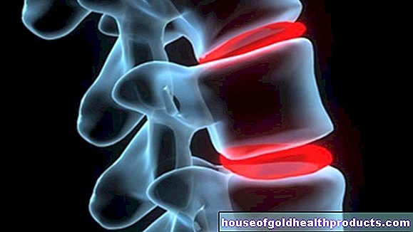
Osteotomy
All content is checked by medical journalists.An osteotomy is the surgical severing of bones or the removal of bone fragments. This procedure is used, for example, to correct the misalignment of a joint. Read all about the process of the osteotomy, when it is done and what the risks are.
What is an osteotomy?
Doctors use an osteotomy to describe the surgical severing of one or more bones, sometimes with the removal of a piece of bone. Doctors often use this procedure if a bone is deformed - then one speaks of a conversion osteotomy or correction osteotomy. The bones are then put back together in the desired position and fixed (osteosynthesis). But this technology is not only used in orthopedics; Dental surgeons also use osteotomies to correct bite misalignments.
When to do an osteotomy
The osteotomy is used to correct misalignments of bones - and teeth. Most osteotomies are performed on the hip, knee, and ankle joints. These joints are particularly stressed, and cartilage wear and tear can develop an unnatural position of the bones in relation to one another over the course of a lifetime. However, misalignments can also be congenital.
Osteotomy: orthopedics
Common orthopedic reasons for a corrective osteotomy include:
- arthrosis
- O- or knock-knees (varus or valgus deformity)
- Different leg lengths
- Misalignments after fractures, for example in the ankle or knee
- Bunion (hallux valgus)
Corrective osteotomies are basically very complex operations. Therefore, one tries to correct bone malpositions first by wearing insoles or splints. If, however, there is no improvement during non-surgical therapy or if the patient has symptoms due to the misalignment that visibly hinder him in everyday life, an operation is necessary.
Osteotomy: dental and maxillofacial surgery
Teeth that are not in the row cannot always be corrected in their position with braces. If they remain in the jaw, inflammation or damage to the neighboring teeth can occur. In such cases, the corresponding tooth or tooth remnant must be removed. Typical dental or maxillofacial surgical reasons for an osteotomy are:
- Teeth or tooth fragments with an atypical location in the jaw
- Wisdom teeth that have too little space in the jaw
- Broken teeth after accidents
- Root remnants left in the jaw before a dental restoration
What do you do with an osteotomy?
In an osteotomy, the surgeon cuts the bone and removes small pieces of bone. In orthopedics, the various techniques differ depending on the affected joint. They are often performed under general anesthesia. In the case of osteotomies on the foot, what is known as conduction anesthesia is usually sufficient, in which the pain-conducting nerves of the foot are switched off in a targeted manner. Before the procedure, the surgeon disinfects the operating area and covers the patient with sterile cloths, leaving out the joint. After the operation, the surgeon places a drainage tube to drain off wound fluid and blood and sutures the layers of muscle, fat and skin.
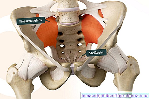
Orthopedic osteotomy of the pelvis
A pelvic osteotomy is necessary when the head of the thigh bone is not correctly positioned in its socket on the pelvic bone. Various procedures are available for the pelvic osteotomy; in adults, the so-called triple osteotomy is usually used. The surgeon uses a bone saw to cut through the three bones of the pelvic ring: the ischium, the pubic bone and the iliac bone. This allows the acetabulum to move freely and the doctor can bring it into the correct position over the head of the thighbone. The surgeon fixes the new position of the bones with screws that remain in the patient's body.
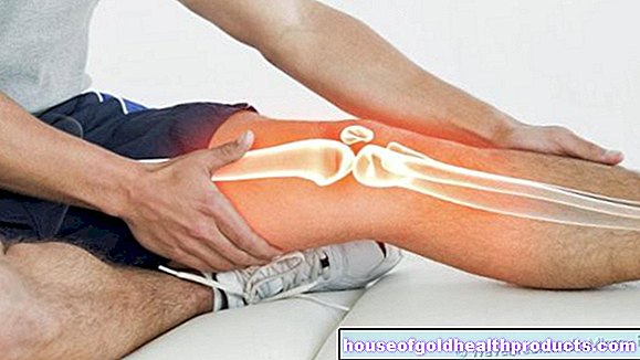
Orthopedic osteotomy of the knee
If loads are unevenly distributed over the knee joint, there will be signs of wear and tear and overloading of the knee joint structures. The surgeon can place the osteotomy on either the thigh bone or the shin bone, depending on which bone is causing the deformity. Here, too, the bone is severed with a bone saw and a bone wedge is cut out. Now the doctor can bring the individual bone parts back into a correct position in relation to one another and fix them with plates or screws.
Orthopedic osteotomy of the foot for bunions
Various surgical procedures are available for treating the bunion: the scarf osteotomy, the chevron osteotomy, the Akin osteotomy and the Weil osteotomy. In many cases, these procedures are also used in combination, as the crooked toe is often based on various misalignments in the individual toe and ankle joints.
With a scarf osteotomy, the surgeon usually makes two small incisions in the skin over which he cuts the shortened ligaments of the metatarsophalangeal joint of the big toe. After exposing the metatarsal bone belonging to the big toe, he cuts it in a z-shape with the bone saw. Now the doctor can move the various bone fragments against each other until they are in the desired position. He then fixes the pieces of bone with a screw so that they do not shift again.
The chevron osteotomy works according to the same principle, but the metatarsal bone is not sawn through in a z-shape, but in a v-shape.
If the bunion is caused by an excess length of the metatarsal bone, a Weil osteotomy is recommended. Here, too, the surgeon cuts the metatarsal bone; he then removes a slice of bone to shorten the metatarsal bone.
If the big toe is still crooked despite the corrective osteotomy in the metatarsal bone, it may be necessary to remove a wedge-shaped piece of bone from the big toe itself. It is known as an Akin osteotomy or base member osteotomy.
Dental and maxillofacial osteotomy
The teeth consist of three areas: the crown, the tooth neck and the root. With the tooth root, they are anchored in recesses in the jawbone (tooth sockets or alveoli). As part of the oral mucosa, the gums cover the necks and roots of the teeth and the tooth sockets.
The osteotomy in the jaw area can be performed on an outpatient basis and, depending on the extent, under local or general anesthesia.
To prevent infections, the doctor first rinses your mouth with an antibacterial mouth rinse. Then he cuts open the gums around the tooth to be removed, folds it to the side and removes parts of the bony tooth sockets with a drill in order to loosen the tooth from its anchorage. To avoid burn damage from friction, the drill is constantly cooled with water.
Once the doctor has loosened the tooth sufficiently, he can lift it out of the tooth pocket with a lever or pliers. He then grinds off sharp bone edges and sutures the gums over the now empty tooth pocket.
What are the risks of an osteotomy?
In general, the osteotomy harbors the following risks, which, however, can generally occur with any operation:
- Bleeding during and after the procedure
- Infections
- Injury to nerves, blood vessels and tendons
- Blood clot formation
- Unaesthetic or painful scarring
- Wound healing disorders
Risks of osteotomy in orthopedics
After the operation, the foot often swells and the movement of the toes is significantly restricted. Lymph drainage is helpful so that the swelling goes down as quickly as possible. Possible complications are also:
- Necrosis of the operated bone (death of cells)
- Slipping or loosening of the inserted screws and plates
- arthrosis
- Another shift in the joint position
- Injury to extensor or flexor tendons with subsequent restriction of movement
- Different leg lengths after osteotomy in the knee and hip area
Risks of osteotomy in dental surgery
In dental and maxillofacial surgery, the following problems can arise during an osteotomy:
- Destruction of dental crowns
- Dislocation of the temporomandibular joint
- Ingestion or inhalation of tooth parts
- Fracture of the lower jaw
- Another shift in the joint position
What do I have to consider after an osteotomy?
In order for an osteotomy to achieve a good result in the long term, your cooperation in the time after the operation is particularly important.
Orthopedic osteotomy
After the osteotomy, you should be absolutely bed rest for two days so that the articular surfaces do not shift again. Then you can get up slowly with physiotherapeutic support. After osteotomies in the foot area, you will receive a special forefoot relief shoe that relieves the recently operated joint as much as possible. As a rule, you can put full weight on your foot with this shoe. It is advisable to wear the forefoot relief shoe for another three to five weeks.
If you experience pain in the wound in the first few days after the operation, your doctor can prescribe an analgesic medication if necessary. The wound dressing is changed every two days. Your doctor will also X-ray the operated joint in the first week after the osteotomy and four weeks after the operation to check that the joint is in the correct position.
Dental surgical osteotomy
Bleeding can often occur within the first 24 hours. It is helpful to cool the operated area with ice packs or cold compresses. Exercise can also trigger bleeding and should be avoided two to three days after the operation. Avoid grainy foods and dairy products and eat soft foods. To prevent infections, daily dental care is important, especially after the operation.Do not use oral irrigators, only use special antibacterial mouthwashes. After the procedure, your dentist will check the wound and the sutures can usually be removed within a week of the osteotomy.
Tags: vaccinations stress drugs