Conduction system
Eva Rudolf-Müller is a freelance writer in the medical team. She studied human medicine and newspaper sciences and has repeatedly worked in both areas - as a doctor in the clinic, as a reviewer, and as a medical journalist for various specialist journals. She is currently working in online journalism, where a wide range of medicine is offered to everyone.
More about the experts All content is checked by medical journalists.The conduction system or excitation conduction system is a system of specifically rebuilt heart muscle fibers. It conducts the regular electrical impulses generated by the so-called pacemaker cells over the entire heart muscle so that it contracts rhythmically. Read everything you need to know about the conduction system!
What is the conduction system?
The conduction system consists of various specialized heart muscle cells that transmit electrical impulses and thus cause the heart muscle to contract rhythmically.
Pacemaker generates electrical impulses
The electrical impulses are generated by so-called pacemaker cells. They are mainly localized in two structures: sinus nodes (the primary pacemaker of the heart) and AV nodes (secondary pacemaker). They both sit in the right atrium and together represent the arousal system.
Normally, the sinus node generates the electrical impulses, which then propagate through the atria to the AV node as the atrium contracts. This is located on the border with the ventricle. From here, the excitation passes through the conduction system to the heart chambers, which then contract.
Like the sinus node, the AV node is capable of spontaneous, automatic impulse generation. However, this only comes into play if the sinus node fails as the primary pacemaker, because the natural frequency of the AV node is 40 to 50 pulses per minute, well below that of the sinus node with around 70 pulses per minute.
Stimulus conduction system: transmission of impulses
The conduction system (excitation conduction system) ensures, as mentioned, that the electrical impulses extend to the entire working muscles of the heart, whereupon the ventricles of the heart contract. The impulses are passed on via defined pathways of specialized heart muscle cells: bundles of His, Tawara thighs and Purkinje fibers.
The bundle of His moves from the AV node through the valve level to the septum between the two main chambers (ventricular septum). There it splits into two branches, which are called the tawara legs (chamber legs). The right leg pulls on the right side of the ventricular septum to the apex of the heart, the left leg on the left side of the septum. Both Tawara legs branch from here to the Purkinje fibers. These run within the working muscles of the heart and ultimately transmit the electrical impulses to the individual muscle cells of the heart chambers, so that they contract. This forces the blood from the left ventricle into the main artery (aorta) and from the right ventricle into the pulmonary artery (pulmonary artery).
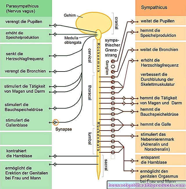
Influence of the nervous system
The conduction system is influenced by the autonomic nervous system (sympathetic and parasympathetic). By stimulating the sympathetic nervous system, the heart rate and the strength of the heart are increased; stimulating the parasympathetic nervous system lowers the heart rate by decreasing the pacemaker rate in the sinus node.
Which problems can arise in the conduction system?
A disturbance can occur at any point in the conduction system. In the case of the atrioventricular block (AV block), for example, the conduction of excitation between the atria and the ventricles is delayed or temporarily interrupted. This occurs most frequently in older people as a result of degenerative changes in the heart, such as coronary heart disease (CHD) or after a heart attack.
The conduction system can also have a disturbance in the area of the tawara thighs (ventricular thighs), which is referred to as intraventricular blockage (thigh block).
Tags: laboratory values healthy workplace teeth


.jpg)