Dermatomyositis
and Sabine Schrör, medical journalistMarian Grosser studied human medicine in Munich.In addition, the doctor, who was interested in many things, dared to make some exciting detours: studying philosophy and art history, working on the radio and, finally, also for a Netdoctor.
More about the expertsSabine Schrör is a freelance writer for the medical team. She studied business administration and public relations in Cologne. As a freelance editor, she has been at home in a wide variety of industries for more than 15 years. Health is one of her favorite subjects.
More about the experts All content is checked by medical journalists.Dermatomyositis (purple disease) is a rare inflammatory muscle disease that also damages the skin. Symptoms include skin changes, muscle pain, and weakness in certain muscle groups. The treatment of dermatomyositis is lengthy and has many side effects, but often significantly improves the quality of life of those affected. Read everything you need to know about the frequency, symptoms, causes, diagnosis and treatment of dermatomyositis.
ICD codes for this disease: ICD codes are internationally recognized codes for medical diagnoses. They can be found, for example, in doctor's letters or on certificates of incapacity for work. M33

Brief overview
- What is dermatomyositis? A rare inflammatory muscle and skin disease that is a rheumatic disease. Also called purple disease because of the often purple skin changes.
- Forms: Juvenile dermatomyositis (in children), adult dermatomyositis (especially in women), paraneoplastic dermatomyositis (in connection with cancer), amyopathic dermatomyositis (skin changes only).
- Symptoms: tiredness, fever, weight loss, later muscle pain and weakness in the shoulder and pelvic area, possibly drooping eyelids or squinting, possibly shortness of breath and swallowing disorders, scaly skin discoloration, swelling and redness in the eye area. Possible complications (such as cardiac arrhythmias, pulmonary fibrosis, kidney inflammation).
- Causes: Autoimmune disease, the causes of which are not fully known. Possibly genetic and triggered by factors such as infections or medication.
- Diagnosis: Interview, physical examination, blood test, muscle biopsy, electromyography (EMG), ultrasound, magnetic resonance tomography, examination for cancerous tumors, if necessary further examinations such as heart ultrasound, lung function test
- Treatment: medication (such as cortisone), muscle training, and physical therapy.
- Prognosis: The treatment can usually significantly alleviate the symptoms or eliminate them completely. Often, however, a slight muscle weakness remains. Complications and accompanying tumor diseases can worsen the prognosis.
Dermatomyositis: description
The term "dermatomyositis" is made up of the Greek words for skin (derma) and muscle (myos). The ending "-itis" means "inflammation". Accordingly, dermatomyositis is an inflammatory disease of muscles and skin. It belongs to the group of inflammatory rheumatic diseases and here to the subgroup of collagenoses (diffuse connective tissue diseases).
The inflammation damages the so-called striated muscles. These are the muscle groups that can be controlled arbitrarily. In addition, symptoms appear on the skin. Blue-violet, scaly discoloration is typical, which is why dermatomyositis is also called "purple disease".
Dermatomyositis: forms
Depending on the age of the patient, the course of the disease and associated diseases, doctors differentiate between different forms of dermatomyositis:
Juvenile dermatomyositis
This is understood to mean dermatomyositis in young people. Children in the seventh and eighth year of life are particularly affected, with girls and boys falling ill at about the same rate.
Juvenile dermatomyositis begins acutely and often affects the gastrointestinal tract as well. An important difference to dermatomyositis in adults: the juvenile variant is never associated with tumor diseases, which is often the case in adult dermatomyositis patients.
Adult dermatomyositis
This is the classic dermatomyositis in adulthood. It predominantly affects women between the ages of 35 and 44 and between 55 and 60 years.
Paraneoplastic dermatomyositis
For many adults with dermatomyositis (20 to 70 percent), the condition is cancer-related, but not directly caused by it. One then speaks of paraneoplastic dermatomyositis. Sometimes it develops parallel to the tumor. In other cases, it precedes or follows cancer.
Paraneoplastic dermatomyositis occurs particularly in the age group from 50 years. Most often, dermatomyositis is associated with the following cancers, depending on gender:
- Women: breast cancer, uterine cancer, ovarian cancer
- Men: lung cancer, prostate cancer, cancer of the digestive organs
Amyopathic dermatomyositis
Doctors speak of amyopathic dermatomyositis when typical skin changes occur, but no muscle inflammation can be detected for six months. About 20 percent of all patients develop this form of dermatomyositis.
Dermatomyositis: frequency
Dermatomyositis is a very rare disease. Every year between 0.6 and 1 in 100,000 adults worldwide develop it. Juvenile dermatomyositis occurs even less frequently - around 0.2 in 100,000 children around the world are affected each year.
Polymyositis
Polymyositis is very similar to dermatomyositis, but does not lead to the typical skin changes. You can find out more about this disease in the article Polymyositis.
Dermatomyositis: symptoms
Dermatomyositis usually begins insidiously and usually develops within three to six months. The first, as yet unspecific, symptoms include fatigue, fever, weakness, and weight loss. Many sufferers also initially suffer from muscle pain that is similar to that of sore muscles. Muscle weakness and skin changes later add to the clinical picture.
It is not always the muscle complaints that are noticeable first and then the skin changes - the sequence of symptoms can vary from patient to patient.
In addition to muscles and skin, other organs are rarely affected by the disease. If the heart or lungs are affected, it can lead to serious complications.
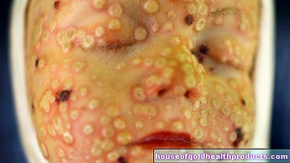
Skin symptoms in dermatomyositis
Skin discoloration (erythema) is characteristic of dermatomyositis. They are usually dark red to blue-violet, but can also present themselves as light red. They usually show up on areas of the skin that are exposed to light - i.e. on the face, neck and arms. The skin can also flake there.
Reddish swollen eyelids are also typical dermatomyositis symptoms - as is a narrow border around the mouth that remains free of discoloration (Shawl's sign).
Other typical signs are redness and raised areas on the skin above the finger joints (Gottron sign) and a thickened nail fold that hurts when pushed back (Keining sign).
Muscle symptoms in dermatomyositis
Muscle pain is typical of the onset of dermatomyositis. They tend to occur under stress. If the disease progresses, increasing muscle weakness develops, which is particularly noticeable near the trunk (proximal), i.e. on the pelvic and shoulder girdles. As a result, those affected find it difficult to perform many movements that involve leg and arm muscles, such as climbing stairs or combing.
The eye muscles can also be affected. This is shown, for example, by a drooping upper eyelid (ptosis) or by squinting (strabismus).
If the throat and respiratory muscles are also affected, swallowing disorders and shortness of breath can occur.
The muscle symptoms of dermatomyositis usually appear symmetrically. If the symptoms only appear on one side of the body, there is probably another disease behind it.
Organ involvement and complications
Dermatomyositis can damage other organs in addition to skin and muscles, which can lead to dangerous complications:
- Heart: Here, dermatomyositis can cause, for example, an inflammation of the pericardium, cardiac insufficiency, a pathological enlargement of the heart muscle (dilated cardiomyopathy) or cardiac arrhythmias.
- Lungs: Lung fibrosis can result from damage to the lung tissue. When dermatomyositis affects the swallowing muscles, the risk of accidentally inhaling food, which can cause pneumonia (aspiration pneumonia), increases.
- Kidneys, gastrointestinal tract: Dermatomyositis can also affect the kidneys or the gastrointestinal tract. Possible consequences are kidney inflammation or intestinal paralysis (intestinal atony).
Overlap Syndrome
In some patients, dermatomyositis occurs together with other systemic immunological diseases, for example systemic lupus erythematosus, systemic sclerosis, Sjögren's syndrome or rheumatoid arthritis.
Dermatomyositis: causes and risk factors
The causes of dermatomyositis are not yet fully understood. Current research suggests that it is an autoimmune disease.
Autoimmune disease
Normally, the immune system can accurately differentiate between the body's own and foreign structures: foreign structures are attacked, but not the body's own. But that is exactly what does not work properly with autoimmune diseases - the immune system suddenly attacks the body's own structures because it mistakenly considers them to be foreign substances.
The underlying mechanisms of many autoimmune diseases are not yet known. This also applies to dermatomyositis. However, scientists suspect that there is a genetic predisposition to the disease. In people with this predisposition, various factors could lead to the outbreak of the disease. Such possible triggers are, for example, infections (for example with Coxsackie, influenza or retroviruses) and medications (for example antimalarials, lipid-lowering agents or anti-inflammatory drugs such as diclofenac):
Certain antibodies then start attacking the blood vessels that supply muscles and skin with oxygen and nutrients. The structures damaged in this way subsequently trigger the typical symptoms of dermatomyositis.
Related to cancer
The likelihood of paraneoplastic dermatomyositis - i.e. dermatomyositis with a connection to a tumor disease - is significantly increased, especially in people aged 50 and over. The exact reason for this is still unclear, although there are some suspicions - such as that a tumor produces toxins that directly damage connective tissue.
In any case, it is known that dermatomyositis often heals after the tumor has been removed, but returns as the cancer progresses.
Dermatomyositis: examinations and diagnosis
The symptoms of dermatomyositis are usually so typical that the doctor can suspect it based on the medical history and physical examination. The disease can be reliably diagnosed with additional examinations. This includes above all blood tests, the examination of muscle samples (muscle biopsy) and the measurement of electrical muscle activity (electromyography, EMG).
Blood test
Certain blood values help diagnose dermatomyositis:
- Muscle enzymes: Elevated levels of muscle enzymes such as creatine kinase (CK), aspartate aminotransferase (AST) and lactate dehydrogenase (LDH) indicate muscle disease or damage.
- C-reactive protein: The C-reactive protein (CRP) is a non-specific inflammatory parameter. So increased values indicate inflammatory processes in the body.
- Blood sedimentation rate (ESR): Increased blood sedimentation can also generally indicate inflammation in the body.
- Auto-antibodies: Antinuclear antibodies (ANAs), Mi-2 antibodies and anti-Jo-1 antibodies attack the body's own tissue and are often (but not always) detectable in dermatomyositis.
While ANAs are found in a number of other autoimmune diseases, the other two auto-antibodies are relatively specific for dermatomyositis (and polymyositis).
Muscle biopsy
Muscle biopsy is the most important test for suspected dermatomyositis. The doctor takes a little muscle tissue for a more detailed analysis: Inflammation and damaged muscle cells can be seen well under the microscope in dermatomyositis.
A biopsy is not necessary if the clinical findings are already clear. This is the case, for example, if the patient shows the typical skin symptoms and demonstrable muscle weakness.
Electromyography (EMG)
In electromyography (EMG), the doctor measures electrical muscle activity with the aid of glued-on electrodes. The results can be used to determine whether the examined muscle is damaged.
Other investigations
Inflamed muscle structures can be visualized using magnetic resonance imaging (MRI) and ultrasound examinations (sonography). MRI is more complex, but also more precise than sonography. Both methods are also used to find suitable sites for an EMG or a biopsy.
Specific examinations are carried out to determine if other organs are involved. These include, for example, cardiac ultrasound (echocardiography), electrocardiography (EKG), chest x-ray (chest x-ray) or a lung function test.
Since dermatomyositis is often associated with cancer (in adults), tumors are also specifically searched for when making a diagnosis.
Dermatomyositis: treatment
Treatment of dermatomyositis usually consists of taking medication, usually for several years. This can counteract the progression of the disease, alleviate symptoms and improve the quality of life for those affected. Muscle training and physiotherapy also make a contribution.
Drug therapy of dermatomyositis
The first choice for dermatomyositis therapy are glucocorticoids (“cortisone”) such as methylprednisolone. In the first four weeks or so, these active ingredients are taken in high doses. The dose is then slowly reduced until, after a few months, it is only necessary to take it every two days, if possible. The gradual reduction is intended to reduce the risk of serious side effects. If the skin symptoms are very pronounced, it can be useful to apply glucocorticoids externally, for example as an ointment.
If the glucocorticoids are insufficient to relieve the symptoms, other immunosuppressants such as azathioprine, cyclophosphamide or methotrexate (MTX) are used. They dampen the immune system to such an extent that it is no longer directed against the body's own structures. However, the defense cannot be completely switched off. And that's a good thing, so that the body continues to be protected from pathogens.
If the mentioned agents cannot improve the dermatomyositis sufficiently, treatment with special antibodies (immunoglobulins) such as rituximab is an option. These fight the immune system exactly where it is acting incorrectly.
Muscle training and physiotherapy for dermatomyositis
Physiotherapy and physical training can support the success of the treatment. Strength and endurance can be increased significantly with the help of a bicycle ergometer or stepper.
It is important to train regularly and to adapt the training intensity to the current state of the disease. Patients should seek advice from their physical therapist or doctor in this regard.
Other measures for dermatomyositis
In the case of paraneoplastic dermatomyositis, the tumor disease must be treated in a targeted manner, for example through surgery, chemotherapy and / or radiation therapy. The dermatomyositis often improves afterwards.
Complications, such as those affecting the heart or lungs, also require special treatment.
Further measures and tips for dermatomyositis:
- UV radiation can worsen the skin changes. Dermatomyositis patients should therefore protect themselves adequately from the sun (sun milk with a high sun protection factor, long trousers, long-sleeved tops, etc.).
- Long-term use of glucocorticoids increases the risk of osteoporosis (weak bones). The doctor can therefore prescribe the intake of calcium and vitamin D tablets as a preventive measure.
- In the acute phase of the disease, dermatomyositis patients should avoid physical activity or stay in bed.
- A balanced diet is also recommended.
Dermatomyositis: disease course and prognosis
In most cases, immunosuppressive therapy can alleviate or even eliminate the symptoms. However, it is possible that an already existing, slight muscle weakness remains. In addition, the complaints can recur at any time.
In some patients, treatment cannot relieve symptoms, but it can at least stop the disease. In others, however, it continues unabated despite treatment.
Risk factors for severe gradients
Old age as well as being male favor a severe course of the disease.The same applies if the heart or lungs are affected. Accompanying cancer is also a risk factor for a severe course of dermatomyositis. Life expectancy can be reduced in such cases.
Tags: toadstool poison plants anatomy pregnancy birth


.jpg)