Macular degeneration
Marian Grosser studied human medicine in Munich. In addition, the doctor, who was interested in many things, dared to make some exciting detours: studying philosophy and art history, working on the radio and, finally, also for a Netdoctor.
More about the experts All content is checked by medical journalists.Macular degeneration is one of the most common causes of adult blindness. The most important part of the retina is destroyed, so that clear vision is no longer possible. In the worst case, there is a risk of extensive blindness. With early treatment, however, the disease can be delayed with medication and minor interventions. Read more about the types and causes of macular degeneration and how to treat them.
ICD codes for this disease: ICD codes are internationally recognized codes for medical diagnoses. They can be found, for example, in doctor's letters or on certificates of incapacity for work. H35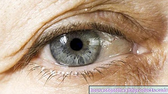
Macular Degeneration: Description
The retina is a special part of the nervous system that lines a large part of the inside of the eyeball. It is responsible for converting light stimuli into nerve impulses: The light hits certain molecules in the photocells of the retina, which generates the nerve impulses. These impulses are passed from the optic nerve to the brain, where they are processed and finally recognized as images.
Retina: structure and function
The retina is made up of many layers made up of different types of nerve cells.The first link in the processing of light signals into nerve impulses are the light sensory cells, the so-called cones and rods. They convert the light stimuli and pass them on to other nerve cells, which in turn are connected to other cells. In this way, the signal is transported via several intermediate stations to the optic nerve and from there to the brain.
The light sensory cells are located in the deepest layer of the retina, so that the light first has to pass through all the other layers. When the light arrives there, a certain cell component, the retinal, changes and splits off small parts ("membrane discs"). It is used up and has to be renewed again.
Disrupted waste removal
The associated retinal pigment epithelium (RPE) is responsible for this reprocessing of the light sensory cells. It transports the resulting waste products away and regenerates the cones and rods.
If this breakdown component is damaged, the metabolic products that accumulate in the retina (e.g. split-off "membrane discs") can no longer be properly transported away. They accumulate and initially destroy the RPE. As a result, the light-sensing cells also perish - macular degeneration occurs.
What happens with macular degeneration?
Macular degeneration is a disease of the retina. However, it does not damage the entire retina, but mainly a specific area. This area is called the macula lutea ("yellow spot"). This is a round, approximately five millimeter large area in the center of the retina, which is contrasted yellowishly with its surroundings due to a special density of light sensory cells.
The light sensory cells of the macula are mainly cones that enable sharp color vision. The other group of light sensory cells (photoreceptors) are the rods. They are responsible for black and white vision in poor light conditions and are therefore particularly important in twilight or at night. Without the yellow spot, one would not be able to read, recognize faces and only perceive the surroundings in outline.
If the macula is destroyed, this results in massive impairment of vision. Since the retina around the yellow spot often remains intact, one does not go completely blind with this disease. Accordingly, in macular degeneration, the edges of the field of vision are still perceived, but not what is fixed in the center of the field of vision.
There are different forms of macular degeneration. By far the most common is age-related macular degeneration, which can appear as a dry or a wet variant. Other forms of macular degeneration caused by genetic defects or other factors are less common.
Age-related macular degeneration (AMD)
The development of age-related or age-related macular degeneration is based - as the name suggests - on aging processes. The destruction of the yellow spot rarely sets in before the age of 60.
In the western industrialized countries, this disease is the most common cause of blindness in old age. It is estimated that around 67 million people in Europe are affected by age-related macular degeneration. Every year around 400,000 new cases occur in Europe. The term “blindness” may be misleading because poor eyesight is retained. In the later stage of the disease, however, one can speak of almost complete blindness.
In poorer countries, age-related macular degeneration is often not the number one cause of blindness. Instead, other eye diseases predominate here, which cannot be adequately treated due to the lack of medical care. Examples are glaucoma (glaucoma) or infectious diseases such as trachoma (a bacterial eye infection).
The dry macular degeneration
About 80 percent of all age-related macular degeneration (AMD) cases are so-called dry macular degeneration (also called dry AMD or non-exudative AMD). In those affected, the inadequately removed waste products of the photoreceptors (especially the so-called lipofuscin) are deposited and in some places form larger associations called "drusen". The extensive damage to the retinal pigment epithelium caused by drusen is also known as "geographic atrophy".
Since dry macular degeneration only progresses slowly over the years, it initially has little effect on eyesight. However, it can turn into wet macular degeneration at any time. This progresses faster.
The wet macular degeneration
Wet macular degeneration (exudative AMD) almost always occurs as a result of dry macular degeneration. What is happening in the eye? The pathological deposits in the retina lead to the destruction of the cells of the retinal pigment epithelium and create gaps in the membranes under the retinal layer. In addition, the blood supply through the choroid is disturbed, which means that the retina is no longer adequately supplied with oxygen in the affected areas.
In response to this, the body images certain messenger substances, so-called growth factors. They stimulate the formation of new small blood vessels - many small vessels sprout from the choroid. The process is called choroidal neovascularization (CNV).
The body would like to counteract the oxygen deficiency. The new vessels also grow through the membrane gaps under the retina, where they actually don't belong. This can cause the retina to peel off, leading to impaired vision and eventually partial or even total blindness. In addition, the walls of the newly formed vessels are not as stable as those of normal blood vessels. Therefore, a little fluid is constantly leaking into the environment, which further promotes retinal detachment. This phenomenon also explains the term “wet macular degeneration”. The small vessels can also tear, causing the retina to bleed.
The wet macular degeneration is much faster and more dangerous than the dry form.
Macular degeneration: symptoms
The macula is the most important area of the retina for vision. If you focus sharply on something with your eyes, this is only possible through the "yellow spot". In the peripheral areas of the field of view, the environment is only perceived as shadowy. Blurred vision from the edge areas around the macula is also important. This is the only way to orientate yourself in space and register movements around you.
Macular degeneration: symptoms in the early stages
In the early stages of macular degeneration, there is often no recognizable visual impairment at all. Although the disease usually affects both eyes in the course of the disease, at the beginning it is often only noticeable in one eye. As a result, the first visual loss in the diseased eye can be compensated for by the still healthy eye. The affected person does not notice anything of the macular degeneration at first. The first symptoms then appear, for example, when reading: The middle of the text may appear slightly blurred, distorted or as if overlaid by a gray shadow.
Macular degeneration in the early stages is often an incidental finding by the ophthalmologist, especially since it does not cause pain.
Macular degeneration: symptoms in the further course
The further the macular degeneration progresses, the more pronounced the symptoms become. Especially when both eyes are affected, i.e. the deficits of one eye can no longer be compensated for by the other eye. In general, the destruction of the "yellow spot" in the middle of the field of vision leads to:
- Decrease in visual acuity
- Decrease in contrast sensitivity
- Decrease in color perception
- Disturbance of adaptation to changing light conditions as well as increased sensitivity to light
- distorted perception of the environment (metamorphopsia)
Due to the fuzzy perception of the central field of vision, those affected can no longer see differences in brightness so well. Contrasts then appear blurred. Because adaptation to changed lighting conditions is also limited, those affected quickly feel dazzled in bright light.
Color vision also suffers, as macular degeneration destroys a large part of the cones in the retina. Those affected increasingly see only in black and white.
The distorted perception (metamorphopsia) is particularly evident when looking at straight lines, such as grid patterns or tile joints. The straight lines suddenly appear curved or wavy. This is how the Macular Degeneration Test works (see below).
When the macular degeneration is well advanced, vision in the center of the field of vision may be completely lost. The patients then only see a light, gray or black spot at this point. In ophthalmology, this spot is called the “central scotoma”.
Macular Degeneration: Causes and Risk Factors
The mechanism that leads to macular degeneration is well known. But why the removal of metabolic products in the eye no longer functions adequately, especially in old age, is still the subject of research.
Confirmed risk factors for macular degeneration are:
- older age: considered to be the most important risk factor. In the 65 to 74 year olds, around 20 percent suffer from AMD, in the 75 to 84 year olds already 35 percent. As society in the western industrialized nations is aging as a whole, macular degeneration is also increasing in frequency.
- Smoking: Nicotine consumption worsens (among other things) the blood circulation in the eye, so that the retina does not get enough oxygen. In addition, metabolic products in the retina are poorly removed by smoking. Those who smoke for many years are therefore more prone to macular degeneration.
- Familial predisposition: As with many diseases, a familial accumulation can also be determined with macular degeneration. Experts suspect that a certain gene constellation makes the risk of (age-related) macular degeneration more likely.
High blood pressure (hypertension), hardening of the arteries (arteriosclerosis) and an increased BMI (body mass index) may also promote macular degeneration. Frequent exposure to sunlight with unprotected eyes is also a suspected risk factor.
Sometimes patients taking the drug chloroquine for malaria prophylaxis or the treatment of inflammatory rheumatic diseases develop macular degeneration over time. However, these are exceptional cases.
A cataract operation - a surgical procedure for cataracts - is increasingly seen as a further cause of the later occurrence of macular degeneration. According to an Australian study, the risk of developing macular degeneration after cataract surgery is five times higher.
Macular degeneration as a result of a genetic defect
Some people develop the typical symptoms of macular degeneration due to a genetic defect, even in childhood and adolescence. Examples of such genetic defects are Best disease (vitelliform macular degeneration) and Stargardt's disease. In the case of Stargardt's disease, the photoreceptors self-destruct.
Macular degeneration as a result of myopia
In rare cases, severe nearsightedness (myopia) can lead to macular degeneration. Myopia is usually the result of an eyeball that is too long. Due to the anatomical disproportion, tension is exerted on the retina. In the long run, this causes the choroid membrane under the macula to thin out, so that at some point the blood supply is no longer sufficient. This creates a wet macular degeneration.
Macular degeneration: examinations and diagnosis
The symptoms of macular degeneration are typical, but are not enough on their own to make a diagnosis. Finally, other diseases of the eye can lead to similar symptoms.
From the age of 55, patients are entitled to regular preventive examinations by an ophthalmologist. In this way, age-related macular degeneration can be detected at an early stage. Various examinations help with this.
Amsler grid
The Amsler grid is named after a Swiss ophthalmologist. It is a recorded, fine-meshed grid with a small, black point in the middle. The patient is half a meter away from the Amsler grid. Now he has to aim at the black point alternately with the right and left eye, whereby the other eye is closed. People with macular degeneration see holes or blurred dark areas in the grid or perceive the grid lines to be distorted and wavy.
The Amsler grid is not a specific macular degeneration test, as it generally shows damage to the retina. However, the grid is used in many ophthalmological practices, especially with older patients, in order to detect age-related macular degeneration at an early stage.
The Amsler grid is also available on the Internet. So if you want, you can first test yourself if you suspect macular degeneration (or retinal damage in general).
Examination of the fundus
The inner surface of the eyeball, which is lined with the retina, is called the fundus of the eye. The doctor can examine the fundus as part of an ophthalmoscopy. He looks into the inside of the eye under illumination through a magnifying glass. Macular degeneration often shows typical structures such as drusen and degenerated, thinned tissue. In the case of wet macular degeneration, spouting vessels, exudates and hemorrhages are also visible.
Usually the fundus is photographed during the ophthalmoscope so that the condition can be compared with later images. In this way, the progression of the disease can be documented.
Fluorescence Angiography (FAG)
Using fluorescence angiography (FAG), macular degeneration can be clearly diagnosed. To do this, a special fluorescent dye is injected into a vein of the patient. It is distributed in the body via the circulatory system and also reaches the retinal vessels. If the fundus of the eye is irradiated with short-wave light, the dye in the vessels glows and makes them visible. For example, newly formed vessels in wet macular degeneration can be easily identified.
Optical coherence tomography (OCT)
For a more precise clarification of macular degeneration, a method is increasingly being used that takes layer images of the retina with weak and harmless laser light. This optical coherence tomography is easier to carry out than fluorescence angiography (nothing has to be injected, for example) and is painless for the patient.
OCT is also used by ophthalmologists to assess the course of the disease and to control further therapy.
Determination of visual acuity
In order to be able to objectively specify the degree of vision loss, the doctor determines the patient's visual acuity (visual acuity). A healthy young person has a visual acuity between 1 and 1.6. In the elderly, it drops to as low as 0.6. However, if there is age-related macular degeneration in the end stage, the visual acuity can drop to below 0.02.
Macular Degeneration: Treatment
The approach to macular degeneration therapy depends on whether it is a wet or dry macular degeneration. Basically, however, there is no treatment that can do anything against the actual cause of the disease. Therefore, the progression of the disease can usually not be prevented in the long term. However, it can be slowed down with medication or certain technical processes and the quality of life of the patient increased. In order to compensate for the loss of vision, at least initially, there are special reading glasses and magnifying glasses.
How to treat dry macular degeneration
In dry macular degeneration, the focus is on the administration of substances that prevent damage to the retinal pigment epithelium in the macula. These include above all zinc and copper oxide as well as so-called antioxidants such as vitamins C and E or beta-carotene. Lutein is a substance that is also found naturally in the macula, where it helps to form the macular pigment. Similar to antioxidants, this natural “dye” protects the photoreceptors in the retina from damage caused by short-wave light or free radicals (aggressive oxygen compounds that can damage cells and genetic material).
In addition, recent studies have shown that the administration of vitamins B6, B12 and folic acid has a positive effect on the course of macular degeneration.
How to treat wet macular degeneration
What makes the wet macular degeneration progress particularly quickly are the new blood vessels. That is why one tries, for example, to destroy the vessels and thus improve visual acuity.
A laser treatment helps with some patients: the diseased vessels are obliterated with laser beams. However, this only works if the vessels are not located directly in the macula. Another disadvantage is that laser macular degeneration treatment also creates scars in the intact tissue, which can impair vision.
In photodynamic therapy, the doctor injects a non-toxic dye into the patient's arm vein. This accumulates in the diseased vessels and makes them sensitive to low-energy laser light. This destroys the vessels in a targeted manner without damaging the surrounding tissue.
Another treatment option for macular degeneration are drugs that contain antibodies (pegaptanib, ranibizumab and aflibercept). They impair the effect of the growth factor VEGF, which stimulates the formation of new blood vessels. The doctor injects the antibody-containing substance directly into the vitreous humor of the eye.
Surgical procedures such as subretinal surgery or retinal rotation (retinal rotation) with displacement of the macula are only useful in rare cases. Some of them are still being tested or further developed.
Therapeutic approaches without guaranteed effectiveness
In the so-called rheophoresis, certain proteins are filtered out of the blood. The method is similar to dialysis and improves the flow properties of the blood. However, their effectiveness in macular degeneration has not yet been proven.
Some people use alternative treatment options for macular degeneration: Acupuncture, for example, can have positive effects in individual cases, especially in dry macular degeneration.
Therapeutic measures that have no guaranteed effectiveness and whose scientific background is questionable should at most be attempted in addition to a treatment with guaranteed effectiveness.
Macular degeneration: disease course and prognosis
How a macular degeneration proceeds varies greatly from person to person. In any case, it is a chronically progressive disease that has not yet been curable.
Dry macular degeneration usually progresses slowly. Sometimes it can even stall for long periods of time. Patients then see no worsening of symptoms for months, sometimes years. However, a complete standstill is very unlikely, although such cases have also been described in isolated cases.
In around 10 percent of cases, a dry macular degeneration eventually turns into a wet macular degeneration. This is progressing very quickly. If one of the diseased vessels ruptures, the resulting hemorrhage into the macula can lead to sudden severe loss of vision. The patients then see considerably worse from one moment to the next.
Macular degeneration: prevention
Since macular degeneration is currently not curable, the disease should be prevented. The most important piece of advice on this: don't smoke! Quitting smoking reduces the risk of macular degeneration in old age.
Tags: sex partnership tcm travel medicine