Veins
Nicole Wendler holds a PhD in biology in the field of oncology and immunology. As a medical editor, author and proofreader, she works for various publishers, for whom she presents complex and extensive medical issues in a simple, concise and logical manner.
More about the experts All content is checked by medical journalists.Veins are blood vessels that carry blood from the body back to the heart. Most of the time they have to work against gravity. Weak veins lead to swollen legs, spider veins or even varicose veins. If these blood vessels are superficial, they are visible under the skin as a bluish path and are well suited for taking blood. Here you can find out everything you need to know about veins!
The way to the heart
The majority of the blood vessels make up 75 percent of the veins. The blood flows from the arterial system into the venules via a network of capillaries. These are the smallest venous vessels with a diameter of 15 to 500 micrometers. They pass into smaller veins (max. Diameter one millimeter), which in turn open into the large body veins (diameter one to ten millimeters). The latter eventually converge in the superior and inferior vena cava, both of which flow into the right atrium.
An important collection point for blood from the abdomen is the portal vein, a vein that brings oxygen-poor but nutrient-rich blood from the abdominal organs to the liver - the central metabolic organ.
However, "used up", ie oxygen-poor, blood does not flow in all veins. The exception is the four pulmonary veins that carry the oxygenated blood in the lungs back to the heart (into the left atrium).
Most of the time, veins are in close proximity to the arteries.
Vein structure
Veins are roughly the same size as arteries, but have a thinner wall (because there is less pressure in them) and therefore a larger lumen. Unlike the arteries, they only have a thin muscle layer in their middle wall layer (media or tunica media). Another difference to the arteries: venous valves are built into many veins (see below).
Superficial and deep veins
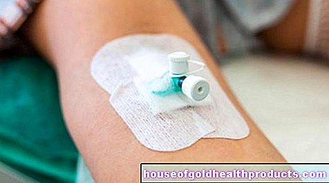
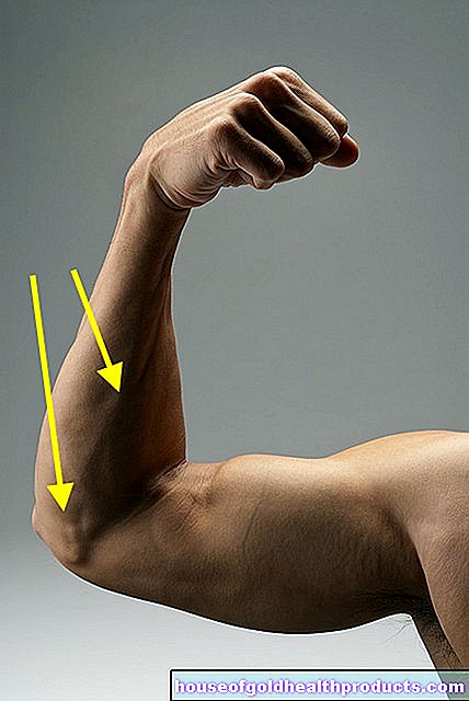
When the blood is drawn, the doctor looks for a superficial vein. They run just under the skin and collect blood from the skin and subcutaneous tissue. Before the blood is drawn, the doctor accumulates the blood in front of the planned puncture site. This works well because the walls of veins are thinner than those of arteries and can expand better. The veins bulge out due to the congestion of blood and are clearly visible due to their blue color. Due to the low pressure inside, the blood flows out of the vein relatively gently after the "prick".
The deep veins run in deeper tissue layers of the body, mostly surrounded by muscles. They contain the majority of the blood volume in the venous system (around 90 percent) and transport the blood from the muscles back to the heart. Superficial and deep veins are in contact with each other via connecting veins.
Veins store a lot of blood
Venous vessels not only carry blood, but are also able to store large amounts of blood. Around five liters of blood flow through an adult's body, more than three liters of it in venous vessels - in an emergency, a crucial reserve to supply vital organs such as the brain and heart. For this reason, in the event of a circulatory collapse, it is important to keep your legs high so that the venous blood can reach the central organs. Even with strenuous physical activity (at work or sport) we are dependent on this extra portion of blood. Without them, no increase in performance would be possible.
Difficult blood transport
The low internal pressure in the venous vessels and the slow blood flow make it difficult to return to the heart. Especially when standing, the venous blood has to be transported against gravity from the bottom to the top. That needs support.
Venous valves
Due to the slow flow of blood against gravity, there is a risk in the veins that the blood will sag, i.e. flow back.To prevent this from happening, venous vessels, especially in the arms and legs, are equipped with venous valves. The pocket-shaped valves made of squamous epithelium only allow blood to flow in one direction (towards the heart). If the blood threatens to flow back, they fold up and prevent the blood from flowing back. In the case of swollen legs or varicose veins, the valves are defective and no longer close completely.
Muscle pump
In addition to the valve system, the skeletal muscles around the veins support their work - but only when we are moving. When sitting or standing for long periods of time, the muscle pump in the legs is hardly active. Then the legs can swell and feel heavy.
Not every vein is equipped with a valve system. For example, veins close to the heart have no valves. They can be supported by the abdominal muscles: Abdominal breathing leads to a drop in pressure in the chest and facilitates the flow of blood into the inferior vena cava. When you exhale, the blood is forced into the right atrium.
Vein training
Spider veins, varicose veins, phlebitis and, in the worst case, a thrombosis or an open leg (ulcus cruris) are typical symptoms of weak veins. The performance of the veins can, however, be trained. When it is warm they expand, when it is cold they contract. Alternating Kneipp baths promote these mechanisms and can alleviate the first symptoms. Exercise, sport and just putting your feet up in between can help. If you sit at your desk a lot, you can train your veins well with simple exercises in between, such as standing on your toes and rocking your feet.
Tags: Diagnosis diet book tip