Knee reflection
Valeria Dahm is a freelance writer in the medical department. She studied medicine at the Technical University of Munich. It is particularly important to her to give the curious reader an insight into the exciting subject area of medicine and at the same time to maintain the content.
More about the experts All content is checked by medical journalists.The inspection of the inner knee joint with a probe is referred to as a knee reflection (knee mirror reflection) or knee arthroscopy. In this way, the doctor can optimally assess the bones, cartilage and the surrounding structures in the knee. At the same time, the doctor can perform operations with the help of additional instruments. Read everything about knee arthroscopy, how it is done and the risks it carries.

What is a knee mirror?
During an arthroscopy of the knee, a so-called arthroscope is inserted into the joint cavity. The arthroscope is a thin tube with a video camera with a light source attached to its end.An additional irrigation and suction device allow an even better examination of the joint. If the doctor detects joint damage, he can treat it in the same session with the help of additional instruments, which are also introduced into the joint cavity through small incisions.
When do you do a knee mirror?
Knee arthroscopy can be used both for diagnosis and for the treatment of degenerative and accident-related injuries. Common reasons are:
- Meniscus damage
- Cartilage damage
- free joint bodies
- Cruciate ligament tears
- Scar tissue
- Inflammation of the synovial membrane
- Gonarthrosis (osteoarthritis of the knee joint)
What do you do with a knee mirror?
As with any arthroscopy, before the actual knee arthroscopy, the doctor asks about the patient's history, explains the risks and benefits of the procedure, carries out a blood test, arranges an anticoagulant drug (heparin) so that no blood clots form in the veins during and after the examination (Thrombosis).
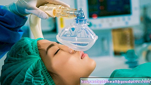
With the help of additional examination methods such as magnetic resonance imaging (MRT), joint damage can be assessed before the arthroscopy and the procedure can be better planned. The skin of the operating area is depilated and carefully disinfected. In contrast to general anesthesia, during which the patient is asleep, with the more frequently used and gentler spinal anesthesia, the pain reliever is injected into the spinal canal so that only the area below the lower back is anesthetized.
Usually a tourniquet is applied. This is an inflatable cuff similar to a blood pressure cuff that is placed around the thigh and inflated to a pressure that is higher than the patient's systolic blood pressure. The tourniquet significantly reduces blood loss.
Now the surgeon opens the joint through a small incision and inserts the arthroscope into the joint cavity. For a better view, this can be filled with a sterile saline solution or carbon monoxide and thus stretched.
Additional instruments can be inserted into the joint via a second and, if necessary, third incision. Cartilage, ligaments and tendons are checked for their strength and functionality with a tactile hook. In the case of a meniscus tear, the injured part can be removed with the help of small scissors or a shaver, a rotating burr. Torn cruciate ligaments are often replaced by the body's own replacement tendons, for example from the patellar ligament (patellar tendon or ligamentum patellare). This type of operation is also called minimally invasive surgery (MIS) and is less stressful than open surgery.
Finally, the arthroscope and the instruments are removed and a small tube (Redon drainage) is temporarily placed in the joint space so that any blood that flows in can be directed to the outside. The incisions are carefully sutured and protected from infections with a bandage and lightly compressed (to reduce the risk of secondary bleeding).
What are the risks of a knee examination?
Knee arthroscopy is a relatively low-risk examination. In rare cases, the arthroscope or other instruments damage the joint itself or surrounding structures such as muscles or ligaments. In addition, vessels and nerves can be injured.
As with any surgical procedure, the knee examination harbors typical surgical risks such as blood clots, intolerance to the anesthetic agent or an infection of both the joint cavity and the remaining skin wounds. Joint effusion, bruising (hematoma) and bleeding may also occur.
What do I have to consider after a knee examination?
Whether the knee examination is carried out on an outpatient or inpatient basis depends, among other things, on the age and health of the patient and the surgery to be expected. Doctors often recommend an inpatient stay of two to three days until the Redon drainage can also be removed.
After a short period of rest, the muscles and ligaments are strengthened and trained with the help of physiotherapy so that the full function of the knee joint can be quickly restored. After a knee examination, the duration of the inability to work is two to six weeks, depending on the physical strain at work and the procedure.
Tags: eyes teenager laboratory values









.jpg)

.jpg)
