Vertebral fracture
Dr. med. Mira Seidel is a freelance writer for the medical team.
More about the experts All content is checked by medical journalists.A vertebral fracture occurs as a result of indirect trauma (e.g. falling from a great height), direct trauma (e.g. fall with direct impact on vertebrae) or osteoporosis. It can express itself through movement-dependent pain. Sometimes a broken vertebra remains symptom-free and then often goes undetected. Depending on the type of fracture, a vertebral fracture is treated conservatively or surgically. Find out more about vertebral fractures here.
ICD codes for this disease: ICD codes are internationally recognized codes for medical diagnoses. They can be found, for example, in doctor's letters or on certificates of incapacity for work. S22T08S32T02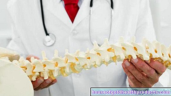
Vertebral fracture: description
The spine consists of a total of seven cervical, twelve thoracic, five lumbar, five sacrum and four to five coccyx vertebrae. Together with a complex ligament and muscle system as well as the intervertebral discs and their characteristic double S shape, the spine is a functional elastic system that can absorb loads.
The vertebral bodies together form the vertebral canal in which the spinal cord (part of the central nervous system) and all of its pathways run. The so-called spinal nerves (peripheral nervous system) originate from the spinal cord and protrude laterally between the vertebrae.
In the event of an overload, the muscle-ligament apparatus can tear and / or a vertebral fracture can occur. This can injure the spinal cord and spinal nerves.
Forms of vertebral fracture
A vertebra consists of a vertebral body, the spinous process and the two transverse processes. Therefore, depending on the location, the vertebral fracture is divided into:
- Vertebral fracture
- Spinous process fracture
- Transverse process fracture
Doctors also differentiate between three different types of fracture, which can run in different directions. This is the Magerl classification, which corresponds to the AO classification (AO = working group for osteosynthesis issues):
- Type A - Compression injuries: This compresses the vertebra, resulting in a cover plate impression or impaction (collapse of the cover plate and base plate of the vertebral body). If the front of the vertebra is compressed, a wedge fracture occurs.
- Type B - Distraction injuries: A torque ruptures the vertebra in the transverse direction. Such injuries occur in the posterior vertebral area. In addition, the intervertebral disc can be torn.
- Type C - Rotational injuries: They occur during a rotation. Longitudinal ligaments and, not infrequently, intervertebral discs are also affected.
Vertebral fractures are also divided into stable and unstable fractures. This is important for the subsequent therapy decision.
Stable vertebral fracture
With a stable vertebral fracture, the soft tissues and the surrounding ligaments remain undamaged. The spinal canal is therefore not narrowed, so that no neurological symptoms occur. The affected person can usually be treated and mobilized at an early stage.
A stable vertebral fracture is, for example, a simple axial compression fracture (type A). Due to compression, the vertebral body is stable against axial forces and also against forces in the flexion direction. 85 percent of all spinal injuries are primarily stable fractures. The following vertebral body fractures are stable fractures:
- Isolated disc injuries
- Isolated vertebral body fracture without disc injury, compression fractures
- Isolated vertebral arch fracture
- Vertebral body fracture with disc injury
Unstable vertebral fracture
An unstable vertebral fracture occurs when the affected spinal column section can be deformed by forces acting from different directions. These include, for example, distraction injuries (type B) and rotational injuries (type C). As soon as the posterior wall of the vertebral body is affected, one speaks of an unstable vertebral fracture, as there is a risk that the spinal cord will be injured by displaced bone fragments. The injury can lead to paraplegia. In the case of unstable fractures, the affected person is restricted in his mobility for a longer period of time.
The following vertebral fractures are unstable:
- Sprained fracture of the vertebrae (usually on the cervical spine)
- Comminuted fracture with damage to the disc tissue and displaced fragments anterior and posterior
- Dislocated fractures with a kink from 25 degrees
- Fractures of the articular processes with gaping spinous processes
- Vertebral arch injuries
Vertebral fracture: symptoms
If a vertebra is broken, local pain typically occurs - regardless of whether the patient is resting, moving or performing strenuous movements. Due to the pain, he usually takes a relieving posture. This can cause the surrounding muscles to tense up (muscle tension).
In the case of cervical vertebrae fractures, those affected often support the head with their hands because of the unstable posture of the head. There may also be a bruise (hematoma) on the back of the neck.
If the vertebral fracture is accompanied by nerve damage, sudden shooting and severe pain (neuropathic pain) as well as painful burning or stabbing (neurogenic pain) can occur. Sensory disturbances (paresthesia) are also possible. In addition, mobility can be restricted in the segment that corresponds to the height of the injury.
Vertebral fracture: causes and risk factors
A vertebral fracture can have various causes. They can be divided into two groups:
Traumatic vertebral fracture
A vertebral fracture is mainly caused by an indirect force, for example a fall from a great height on the legs (chain fracture), on the buttocks or the head. Direct traumas such as a blow to the spine or an open vertebral fracture after a gunshot wound are extremely rare. But even with simple minor traumas such as somersault on the exercise mat or a stump in the parking lot, it can lead to a severe spinal fracture with serious consequences.
In general, the transitions between the cervical spine and thoracic spine, between thoracic spine and lumbar spine and between lumbar spine and sacrum are particularly at risk of injury. About half of all vertebral fractures affect the transition between the thoracic spine and the lumbar spine. The following typical situations can lead to spinal trauma:
- Pelvic belt injuries ("seat belt injuries") can cause a vertebral fracture along with injuries to the abdomen.
- When falling from a great height, a fracture of the heel bone often occurs along with a fracture of the thoracic and lumbar spine.
- Intervertebral discs and ligament structures can tear if rapid body movement is stopped abruptly (deceleration trauma).
Spontaneous fracture of the vertebra
If a vertebral fracture occurs without a corresponding accident, other causes must be considered. Osteoporosis (bone loss) plays a particularly important role in the elderly. The bone loses bone mass and becomes unstable. Often a small amount of force is enough to break the vertebra.
A vertebral fracture caused by osteoporosis is also known as a "sintering fracture". The base and cover plates break in as a so-called fish vertebra or the front wall of the vertebral body as a so-called wedge vortex. This happens especially often in the lower thoracic spine and upper lumbar spine. If old people fall on their face, they often suffer a dens fracture - a type of neck fracture (dens = thorn-like extension of the second cervical vertebra).
Apart from osteoporosis, the following diseases can also lead to an unexpected vertebral fracture in the case of minor minor trauma:
- Bone metastases, bone tumors
- ankylosing spondylitis
- Plasmacytoma (multiple myeloma - a type of blood cancer)
- Inflammation of the vertebrae (spondylitis)
Vertebral fracture: examinations and diagnosis
The specialist responsible for suspected vertebral fractures is a doctor specializing in orthopedics and trauma surgery. He will first ask you about a previous accident and your medical history (anamnesis). Possible questions include:
- Did you have an accident? What happened there?
- Was there any direct or indirect trauma?
- Do you have pain? If so, in which area and with which movements?
- Have there been previous injuries or previous damage?
- Have you had any complaints before?
- Do you have numbness in your arms or legs?
- Have you also had gastrointestinal complaints, difficulty urinating or swallowing problems?
The questions about numbness, swallowing disorders, etc. stem from the fact that around ten percent of all spinal injuries also injure nerves, which can cause such symptoms. In addition, the underlying trauma is usually severe, which can also affect the kidneys and spleen, for example.
Clinical examination
During the clinical examination, the doctor checks whether it is possible to walk or stand. It also tests the patient's general mobility. Next, the cranial nerves, sensitivity and motor skills are checked to see if there are any neurological deficits. In addition, the doctor checks whether there is tension or hardening in the muscle (muscle tension) or a wry neck (torticollis).
Imaging procedures
An X-ray examination in two planes is an important part of the diagnosis of the vertebral fracture. Function recordings are also made. They allow a precise assessment of whether intervertebral discs or ligaments were injured as well. In addition, the distances between the spinous processes of the vertebrae, the vertebral body cavities and the shape of the vertebrae are assessed.
Computed tomography (CT) is particularly suitable as an imaging method for areas that are difficult to see. This is especially true for the transition area between the cervical and thoracic spine. Injuries in this area can be precisely assessed using CT. If there are nerve failures, a CT is always done.
Magnetic resonance imaging (MRI) is usually not required for acute injuries. It is only used when the spinal cord and intervertebral discs could also be injured.
Vertebral fracture: treatment
In principle, vertebral fracture therapy can be carried out both conservatively and surgically. Which method is most suitable in each individual case depends on the type of injury (such as stable or unstable fracture) and also on the age of the patient.
Vertebral Fracture Treatment: Conservative
A stable fracture is usually treated conservatively. The patient is advised to take it easy and keep bed rest until the pain has improved. However, in some cases, the changed shape of the broken vertebral body can bend the spine. A sharp curvature can lead to permanent discomfort. In the case of a curvature of 20 degrees or more in the thoracic and lumbar spine area, surgery is therefore usually carried out.
In the event of a stable fracture of the cervical spine, it can be realigned with an extension (Crutchfield) under X-ray control - the vertebral joints are stretched in the axial direction. The cervical spine is then immobilized with a soft collar (Schanz tie), a hard collar (Philadelphia tie), a Minerva cast or a halo fixator.
In the conservative therapy of vertebral fractures in the thoracic and lumbar spine, a three-point corset or a plaster of paris (plastic) corset is used.
Vertebral fracture treatment: Surgical
An unstable vertebral hernia is usually operated on because there is always the risk that the spinal cord will be injured or already injured. The aim of surgical treatment is to quickly realign and stabilize the spine in order to relieve the pressure on the nerves as quickly as possible. This also applies to complete paraplegia - even if it is not possible to assess whether there will be any improvement after an operation. It is always difficult to predict to what extent the spinal cord of the affected person is damaged.
There are various surgical procedures that can be considered for a vertebral fracture: If nerves are also affected, a so-called laminectomy is performed. The surgeon removes parts of one or more vertebral bodies.
For spontaneous fractures, such as those caused by osteoporosis, either a kyphoplasty or a vertebroplasty is performed.
In principle, two procedures are used for traumatic fractures: osteosynthesis or spinal fusion.
Vertebral hernia surgery: kyphoplasty
Kyphoplasty is a minimally invasive method in which the collapsed vertebral body is straightened up again with a balloon. The surgeon then stabilizes the height of the vertebra by injecting cement.
Vertebral hernia surgery: vertebroplasty
Vertebroplasty is also a minimally invasive method to stabilize the broken vertebral body. Here, too, cement is injected into the vertebral body.
Vertebral fracture surgery: osteosynthesis
With osteosynthesis, the bone fracture is screwed or flattened. A fracture of the dens (thorn-like extension of the second cervical vertebra) or a bilateral fracture of the vertebral arch is usually screwed. Fractures of the thoracic and lumbar spine are fixed over several segments (internal fixator).
Vertebral hernia surgery: spinal fusion
In a spondylodesis treatment (stiffening operation), two or more vertebrae are stiffened with a bone chip or plate. This procedure is usually an option for injuries to the ligaments and discs of the cervical spine. Plates are attached to the cervical spine from the front and back.
If the spine is arched forwards more than 20 degrees due to a compression fracture in the thoracic and lumbar spine area, the vertebral fracture is fused from the front and back. Distraction and torsion injuries to the thoracic and lumbar spine are also stiffened from both sides.
Vertebral fracture: disease course and prognosis
The course of the disease and the prognosis for a vertebral fracture are usually good. However, it plays a major role here whether nerve tissue has been injured. In addition, even after the trauma, there is still the risk that the spinal canal will be narrowed or that neighboring segments will change degeneratively. The following long-term effects can occur after spinal injuries:
- Static disorder: After the vertebral fracture has healed, orthopedic problems with regard to statics can arise.
- Spinal Cord Lesion: All vertebral injuries are at risk of injury to the spinal cord or nerve roots. In the extreme case, paraplegia occurs.
- Post-traumatic kyphosis: If the vertebrae collapse from the front, the backward convex curvature of the spine can intensify. In the thoracic spine, the curvature in the chest area can increase ("widow's hump") and decrease in the lumbar spine area.
- Post-traumatic scoliosis: A lateral curvature of the spine (scoliosis) occurs when the side edges become lower. This scoliosis is short-arched. The statics are influenced by the trunk overhanging and the intervertebral discs above and below being subjected to increased stress.
- Schipper's disease: During heavy physical work such as "shoveling" the spinous processes of vertebrae can break, especially of the seventh cervical or first thoracic vertebra. However, this does not cause any major discomfort.
Vertebral fracture: healing time
The time it takes to heal a vertebral fracture depends on how severe the injuries are. A stable vertebral fracture usually becomes firm again in a few weeks to months without shifting any further. Affected people can get up immediately or after about three weeks, depending on the pain. However, an unstable vertebral fracture can shift further, which creates the risk of compressing the spinal cord and resulting in paraplegia.
Tags: skin care first aid sports fitness


.jpg)