Chest drain
Updated on All content is checked by medical journalists.Chest drainage uses a tube to suck air, blood, or other fluid out of the chest. This is especially necessary after accidents, major operations or malignant diseases in the chest cavity. Read everything you need to know about the chest tube - how it works, when it is performed and what the risks are.

What is a chest tube?
Strictly speaking, the chest tube refers to several types of drainage of fluid or air from the chest. In everyday parlance, however, doctors usually understand a thoracic drainage to be the so-called pleural drainage. Here, the tube lies in the space between the pleura and the pleura, in the so-called pleural space.
Other anatomical spaces in the chest that doctors often have to drain are the mediastinum in the middle between the two lungs - here are important organs such as the heart and the esophagus and trachea - and the pericardium. Depending on what the drainage is from, it is called mediastinal drainage, pericardial drainage or - as explained above - pleural drainage.
When do you perform a chest tube?
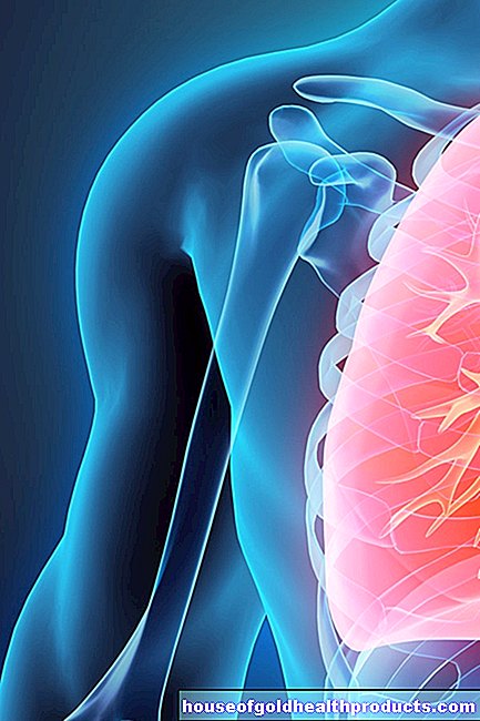
A thoracic drainage in the sense of a pleural drainage is always used when air, blood or other fluid (e.g. pus or lymph fluid) collects in the pleural space. This accumulation of air or fluid prevents the lungs from expanding and can therefore cause severe shortness of breath. In severe cases, it also pinches the large veins and arteries in the heart. The increase in pressure inside the chest is a life-threatening situation and can only be prevented by drainage. The following diseases make chest drainage necessary:
Pneumothorax and tension pneumothorax
If the lungs are injured by accidents to the ribs - often for no apparent cause in slim people - or when the lungs are injured in major open thoracic operations, air often escapes into the pleural space (pneumothorax). In addition to pain, the patient usually also feels shortness of breath and possibly palpitations.
It can be life-threatening if the air enters the pleural space when inhaling, but cannot flow out again when exhaling. The reason can be, for example, destroyed tissue that seals the injury like a valve during exhalation. If left untreated, the pressure in the chest increases until the heart can no longer work. With this so-called tension pneumothorax (also known as "valve pneumothorax"), the doctor must urgently put a chest drainage (read: pleural drainage) in order to save the patient's life.
Pleural effusion
A pleural effusion is a build-up of fluid in the pleural space. The cause is often a weak heart muscle (cardiac insufficiency, heart failure): the heart can no longer pump the blood in the lungs, which increases the pressure in the pulmonary vessels. As a result, some of the water contained seeps into the lung tissue and finally into the pleura (pleural space).
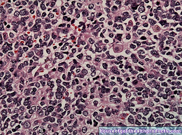
In addition to cardiac insufficiency, cancer-free pleural effusions, such as a malignant tumor in the pleura, often also produce. It is therefore important to examine the fluid drained from the pleural space specifically for malignant cells.
If the pleural effusion usually develops slowly, a chest drain is not the first choice: First, the doctor tries to draw off the liquid with a larger syringe. However, if this measure does not have a lasting effect, he must insert a chest tube.
Hemothorax
The hemothorax is a special form of pleural effusion. Blood collects in the pleural space - for example when there is heavy bleeding due to an accident or - less often - from lung cancer through the rupture of large blood vessels in the chest area. Patients are in pain, their breathing is acutely impaired, and the body can suffer critical blood loss in a short period of time.
Here, too, the doctor has to insert a chest tube as quickly as possible in order to relieve the lung tissue. If this is not enough (e.g. due to a bleeding disorder in the person affected) and the bleeding continues, the only way to save the patient's life is to have the doctor open the chest and stop the bleeding in a targeted manner.
Pleural empyema
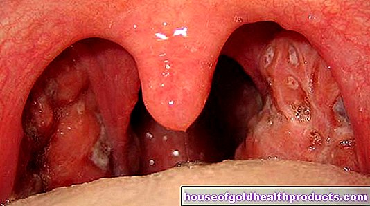
During severe infections, such as after surgery or pneumonia, large amounts of pus can accumulate within the pleural space. Doctors call this a pleural empyema. To remove the pus, the doctor also puts a chest tube here. The chest is then rinsed with saline solution through the tube until the inflammation has healed after a few days - with the accompanying administration of antibiotics.
What do you do with a chest tube?
Before starting a chest drain, it is important to find out exactly where the accumulated air or fluid is with the help of X-rays (taken in several planes) or computed tomography. In emergency medicine, however, these technical examinations are sometimes not available, which is why the emergency doctor has to rely on indicative symptoms and simpler examinations such as tapping and listening to the chest.
Puncture sites for chest drainage
There are two more precisely defined locations available at which the chest can be punctured for the placement of a thoracic drainage:
- Monaldi position: in the middle of the collarbone between the second and third ribs
- Bülau position: laterally below the armpit between the fourth and fifth or fifth and sixth ribs
First of all, doctors try to place a chest tube in the Bülau position and to push the drainage tube further forward in the pleural space. However, the Monaldi position is often the better choice, especially for permanently lying patients who have suffered a pneumothorax in the upper area of the lungs. This is because the air rising up in the pleural space can be better absorbed.
Prepare and put on
First, the doctor must thoroughly cleanse, disinfect and numb the area of skin around the planned drainage site with a local pain reliever. Then he pricks the skin over the intercostal space with a scalpel. Through this small opening he pushes the muscles between the ribs apart with scissors or sometimes with his bare finger and opens up the pleura. It does this along the top edge of a rib, since nerves and blood vessels run along the bottom edge that can be very easily injured.
In the case of tension pneumothorax, after the pleural space has been successfully opened, a loud hissing sound can often be heard when the pent-up air escapes. But even blood or watery-serous fluid can then drain away. After this first evacuation, the doctor uses a metal rod to insert a plastic tube further into the pleural space, which permanently drains air or secretion. This is attached to the skin with a thread using a special technique known as the purse-string suture. This is to prevent the hose from slipping and also to seal the wound well from the outside.
Finally, the doctor connects the end of the hose with a device that creates a constant, slight negative pressure in the hose system. As a result, newly accumulating fluid or air is sucked out of the pleural space immediately. The thoracic drainage creates space for the lungs again.
Removal of the chest tube
If, after a few days, no more secretions collect in the collecting container, no more air is sucked out and no more liquid or air can be seen in the X-ray image, the doctor can pull out the chest drain. To do this, he asks the patient to breathe in deeply and then breathe out forcefully. He quickly pulls out the hose. The purse-string suture is then pulled together tightly and knotted again so that no more air enters the pleural space.
What are the risks of a chest drain?
The severing of the intercostal muscles and the pleura in particular harbors some risks. Since an artery, a vein and a nerve run along the lower edge of a rib, there is a risk that the doctor will injure them. Possible consequences are bleeding or permanent abnormal sensations or numbness in the skin area supplied by the corresponding nerve.
When inserting the actual thoracic drainage tube, the metal guide rod can injure organs in the immediate vicinity, especially under special anatomical conditions. These include the heart, lungs, esophagus and trachea, liver and diaphragm, but also the major artery. If such structures are injured while the chest tube is being placed, emergency surgery is often performed immediately.
Bacteria or other pathogens can penetrate the pleural space through the wound created by the thoracic drainage system and cause infections there.
What do I have to consider after a chest tube?
If doctors have placed a chest tube on you, you should avoid pulling on the tube so that it does not accidentally slip out of your chest.
Also, look out for pain, redness, or swelling in the area of the wound. Let your doctor know if you experience these symptoms - they could indicate the onset of infections.
If you notice that the pleural drainage suddenly produces much more secretion, especially blood, than before, inform your doctor immediately! This is an indication of a new bleeding. You should also inform the doctor in the event of sudden shortness of breath or rapid heartbeat.
If your doctor tells you he wants to pull the chest tube, you can ask about a pain reliever as a preventative measure if you are worried about pain when removing the tube. This ensures that you do not find it uncomfortable to pull out the chest tube.
Tags: interview sleep alcohol drugs


.jpg)